News

Erin Bush Receives Award

Erin Bush with Department of Systems Biology Assistant Professor Peter Sims and College of Physicians & Sciences Dean Lee Goldman. (Photo: Amelia Panico)

The Department of Systems Biology is proud to congratulate Erin Bush on being selected for the Columbia University College of Physicians & Surgeons 2016 Officer of Research Award. The award is one of six given annually to recognize select staff members for their outstanding contributions in the workplace. Recipients of the 2016 were recognized in a ceremony that took place at Columbia University Medical Center on January 12, 2017.

Erin is a staff associate in the JP Sulzberger Columbia Genome Center and a sequencing specialist working in the laboratories of Peter Sims and Andrea Califano. She has been helping to develop new next-generation sequencing techniques, focusing on low input and single cell DNA and RNA library preparation and testing. As the CUMC Newsroom reports:

Noted for her technical skill and professionalism, Ms. Bush was honored for her work in the Department of Systems Biology, where her time and expertise are split among three laboratories. At the Sulzberger Columbia Genome Center, her efforts boosted efficiency at the sequencing core facility and enabled the core’s expansion. More recently, she helped develop an RNA-sequencing technology with the Califano and Sims labs that allows researchers to screen drugs for genetic effects at low cost and high throughput. The new technology is a promising tool for disease research and precision medicine and has led to multimillion-dollar federal grants within the department.

The P&S Annual Awards recognize one employee each in the categories of Management, Administration, Research, Clerical & Technical, Diversity, and Community Service.

Congratulations, Erin!

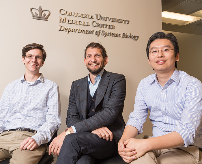
Peter Sims, Sagi Shapira, and Harris Wang

Assistant Professors Peter Sims, Sagi Shapira, and Harris Wang recently moved into a new Department of Systems Biology laboratory space designed to facilitate the development of new technologies for biological and biomedical research. Photo: Lynn Saville.

The Columbia University Department of Systems Biology has opened a new experimental research hub focused on biotechnology development. Occupying one and a half floors in the Mary Woodard Lasker Biomedical Research Building at Columbia University Medical Center, the facility will promote the design and implementation of new experimental methods for the study and engineering of biological systems. It will also enable a substantial expansion of Columbia’s next-generation genome sequencing capabilities.

The first occupants of the new facility are the laboratories of Department of Systems Biology Assistant Professors Sagi Shapira, Peter Sims, and Harris Wang, along with the Genome Sequencing and Analysis Center of the JP Sulzberger Columbia Genome Center. The community is slated to grow, as currently unoccupied space will soon accommodate additional Columbia University faculty labs that are also developing new biotechnologies.

“Technology drives science,” says Department of Systems Biology Chair Andrea Califano, “and the ability to design new technologies can make it possible to answer questions that no one else can. By bringing technology-focused investigators and the Genome Center’s sequencing infrastructure together in the same physical location, our goal is that the new Lasker facility will give the Department of Systems Biology — and the entire Columbia University research community — access to unique applications for biological and biomedical research.” 

Alex Lachmann
Alex Lachmann during his presentation to the RNA-Seq "boot camp."

In June 2015, the Columbia University Department of Systems Biology held a five-part lecture series focusing on advanced applications of RNA-Seq in biological research. The talks covered topics such as the use of RNA-Seq for studying heterogeneity among single cells, RNA-Seq experimental design, statistical approaches for analyzing RNA-Seq data, and the utilization of RNA-Seq for the prediction of molecular interaction networks. The speakers and organizers have compiled a list of lecture notes and study materials for those wishing to learn more. Click on the links below for more information.

Tracking clones

After identifying T cell clones that react against donated kidney tissue in vitro, new computational methods developed in Yufeng Shen's Lab are used to track their frequency following organ transplant. The findings can help to predict transplant rejection or tolerance.

When a patient receives a kidney transplant, a battle often ensues. In many cases, the recipient’s immune system identifies the transplanted kidney as a foreign invader and mounts an aggressive T cell response to eliminate it, leading to a variety of destructive side effects. To minimize complications, many transplant recipients receive drugs that suppress the immune response. These have their own consequences, however, as they can lead to increased risk of infections. For these reasons, scientists have been working to gain a better understanding of the biological mechanisms that determine transplant tolerance and rejection. This knowledge could potentially improve physicians’ ability to predict the viability of an organ transplant and to provide the best approach to immunosuppression therapy based on individual patients’ immune system profiles.

Yufeng Shen, an assistant professor in the Columbia University Department of Systems Biology and JP Sulzberger Columbia Genome Center, together with Megan Sykes, director of the Columbia Center for Translational Immunology at the Columbia University College of Physicians and Surgeons, recently took an encouraging step toward this goal. In a paper published in Science Translational Medicine, they report that the deletion of specific donor-resistant T cell clones in the transplant recipient can support tolerance of a new kidney. Critical to this discovery was the development of a new computational genomics approach by the Shen Lab, which makes it possible to track how frequently rare T cell clones develop and how their frequencies change following transplantation. The paper suggests both a general strategy for understanding the causes of transplant rejection and a means of identifying biomarkers for predicting how well a transplant recipient will tolerate a new kidney.

Hynek WichterleThe JP Sulzberger Columbia Genome Center is pleased to announce that Hynek Wichterle has been appointed as associate director. In this role he will advise on stem cell related projects and coordinate interactions between Columbia Stem Cell Facility and the Columbia Genome Center's High-Throughput Screening Facility.

In addition to his position at the Columbia Genome Center, Dr. Wichterle is also an associate professor holding a joint appointment in the Departments of Pathology & Cell Biology and Neuroscience (in Neurology) at Columbia University Medical Center. He received his MS degree from Charles University in Prague and his PhD degree from The Rockefeller University. He trained at Columbia University, where he became assistant professor in 2004 and associate professor in 2012. He serves as a co-director of the Columbia Stem Cell Initiative and as a Vice-Chief of the Division of Regenerative Medicine in the Department of Rehabilitation & Regenerative Medicine.

Dr. Wichterle developed groundbreaking methods for producing spinal cord neurons from pluripotent embryonic stem cells in a culture dish. The process faithfully recapitulates normal embryonic development, providing a unique opportunity to study and experimentally probe nerve cells in a controlled environment outside of the embryo. He is using the system to decode transcriptional programs that control genes important for neuronal differentiation and function. His lab also capitalizes on the unlimited source of spinal neurons to study motor neuron degenerative diseases, such as amyotrophic lateral sclerosis (ALS or Lou Gehrig’s disease), with the goal of discovering new drugs for these currently untreatable, devastating conditions.

Fluidigm C1 Single-Cell Plate

At the core of the Fluidigm C1 Single-Cell Auto Prep System is a 96-well plate containing microfluidics. After individual cells are isolated in their own wells, the device amplifies their cDNA for genome-wide gene expression profiling. Scientists at the Columbia Genome Center are developing methods for addressing the technical and analytical challenges of single-cell RNA sequencing, and have begun generating some exciting data.

Since the invention of the first microscope, a procession of new technologies has enabled scientists to study individual cells at increasingly fine levels of detail. The last two years have witnessed an important next stage in this evolution, with the arrival of the first devices for genetically profiling single cells on a genome-wide scale.

The first commercial product in this field is the Fluidigm C1 Single-Cell Auto Prep System, which uses microfluidics to isolate single cells and offers the ability to generate gene expression profiles for up to 96 cells at a time. But because of the novelty of the technology and the inherent difficulties of working with single cells, it has presented a number of technical challenges for researchers interested in exploring biology at this level.

Now, scientists at the JP Sulzberger Columbia Genome Center led by Assistant Professors Peter Sims and Yufeng Shen have developed an experimental and computational pipeline that optimizes the C1’s capabilities. And even as they work to solve some of the challenges that are inherent to single-cell research, their approach has begun generating some exciting data for studying genetics in a variety of cell types.

Illumina NextSeq 500 at Columbia University

As genome sequencing technologies evolve, the JP Sulzberger Columbia Genome Center continues to provide the Columbia University biological and biomedical research community access to state-of-the-art tools. In its most recent acquisition, the Genome Center has just installed two Illumina NextSeq 500 sequencers. The NextSeq 500 is a flexible and efficient desktop sequencer that offers powerful high-throughput sequencing capabilities.

Columbia investigators who are experienced with the Illumina next-generation sequencing platform can now schedule time to use the NextSeq 500 for their own research. 

Peter SimsPeter Sims, an assistant professor in the Columbia University Department of Systems Biology, has been named Associate Director for Novel Technologies at the JP Sulzberger Columbia Genome Center. In this role he will devise, direct, and implement strategies for incorporating new high-throughput experimental methods into the research done at the Genome Center.

Trained as a physical chemist, Dr. Sims has been developing a number of innovative technologies for studying single cells in a high-throughput setting. Using a type of microfluidics called soft lithography, his laboratory has designed a method for creating arrays composed of wells just tens of microns in diameter, small enough to isolate and perform high-throughput experiments on individual cells.  

Appointing Dr. Sims to his new role will enable the Columbia Genome Center to develop a variety of new applications that will benefit researchers across the Columbia University community. 

Imaging synapses

In a test of the Columbia Genome Center's high-content microscopy system, computational image analysis confirmed a high degree of colocalization of green fluorescent protein-labeled synapsin and the dye FM4-64. The researchers plan a high-throughput screen to identify fluroescent small molecules capable of targeting synapses.

Synapses mediate communication between neurons in the brain, making them critical components for neurological activity. Research has shown that synaptic loss and dysfunction play roles in a number of debilitating brain disorders — including Alzheimer’s disease, major depressive disorder, and autism — but currently no effective method exists for identifying and imaging individual synapses in living human brains. Being able to locate and quantify synapses in patients could greatly improve the diagnosis and monitoring of disease, and potentially offer new approaches for treatment.

Clarissa Waites, an assistant professor of pathology and cell biology at the Columbia University College of Physicians & Surgeons, and Dalibor Sames in the Columbia University Department of Chemistry, have recently embarked on a collaboration with the Columbia Genome Center High-Throughput Screening Facility with the goal of identifying small fluorescent molecules that can selectively localize to synapses. If successful, this project could for the first time provide a method for targeting and imaging synapses in the living human brain.

Rabadan, Nature Genetics

An analysis of all gene mutations in nearly 140 brain tumors has uncovered most of the genes responsible for driving glioblastoma. The analysis found 18 new driver genes (labeled red), never before implicated in glioblastoma and correctly identified the 15 previously known driver genes (labeled blue). The graphs show mutated genes that are commonly found in varying numbers in glioblastoma (left), that frequently contain insertions (middle), and that frequently contain deletions (right). Genes represented by blue dots in the graphs were statistically most likely to be driver genes.

A team of Columbia University Medical Center researchers has identified 18 new genes responsible for driving glioblastoma multiforme, the most common—and most aggressive—form of brain cancer in adults. The study was published August 5, 2013, in the journal Nature Genetics.

The Columbia team used a combination of high-throughput DNA sequencing and a new method of statistical analysis developed by co-author Raul Rabadan, an assistant professor in the Department of Systems Biology, to generate a short list of candidate gene mutations that were highly likely to drive cancer, as opposed to mutations that have no effect.

Considering these results along with a previous study this group conducted, Rabadan and collaborators Antonio Iavarone and Anna Lasorella point out that approximately 15% of glioblastomas could now be targeted with drugs that have already been approved by the FDA. As Lasorella remarks in an article for the CUMC Newsroom, “There is no reason why these patients couldn’t receive these drugs now in clinical trials.”

Archive