New NIH Grant to Accelerate Commercialization of Single-Cell Analysis Platform
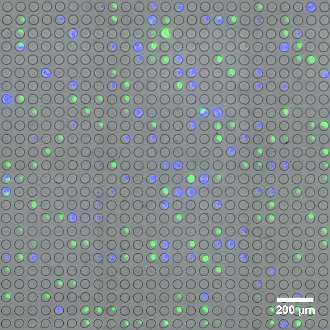
Microwell array flow cell device loaded with fluorescently-labeled live cells. (Image provided by the Sims Lab.)
The field of single-cell RNA sequencing is moving at a fast clip. Adding to its rapid advance is a novel platform for linking optical imaging with high-throughput single-cell sequencing devised by researchers in the Sims Lab at Columbia’s Department of Systems Biology.
The new technology, developed by Peter Sims, PhD , assistant professor of systems biology, and postdoctoral research scientist, Jinzhou Yuan, PhD , enables live cell imaging and RNA sequencing simultaneously of the same individual cell on a large scale and at low cost. Jointly awarded a $1.5 million grant funded by the National Institutes of Health’s SBIR program, the Sims Lab and Cell Microsystems are collaborating to build and test the device, with the goal of bringing to market a fully-integrated system capable of imaging thousands of single cells and preparing them for genomic analysis.
The proposed system will integrate Cell Microsystems’ proprietary CellRaft Technology with the Sims Lab’s novel approaches to tracking single cells with so-called optical barcodes.
In single-cell RNA sequencing, “State-of-the-art technology now allows us to routinely process thousands of individual cells and obtain their genome-wide mRNA expression profiles,” notes Dr. Yuan. “However, cells that share similar expression profiles at the mRNA level may be distinct from each other based on features obtainable by optical microscopy, such as morphology, motility, and fluorescent labels. A more comprehensive description of each cell obtained by both imaging and sequencing may help us further refine cell type and state.”