Immunology ×

News

Breast cancer cells

A histological slide of cancerous breast tissue. The pink "riverways" are normal connective tissue while areas stained blue are cancer cells. (Source: National Cancer Institute)

Investigators at Columbia University Medical Center and the Icahn School of Medicine at Mount Sinai have discovered a molecular signaling mechanism that drives a specific type of highly aggressive breast cancer. As reported in a paper in Genes & Development, a team led by Jose Silva and Andrea Califano determined that the gene STAT3 is a master regulator of breast tumors lacking hormone receptors but testing positive for human epidermal growth receptor 2 (HR-/HER2+). The researchers also characterized a pathway including IL-6, JAK2, STAT3, and S100A8/9 — genes already known to play important roles within the immune response — as being essential for the survival of HR-/HER2+ cancer cells. Additional tests showed that disrupting this pathway severely limits the ability of these cells to survive.

These findings are particularly exciting because the pathway the researchers identified contains multiple targets for which known FDA-approved drugs exist. The paper reports that when these drugs were tested in disease models, the cancer cells showed a dramatic response, suggesting promising strategies for the treatment of the HR-/HER2+ cancer subtype. A clinical trial is now underway to investigate the effects of these approaches in humans.

Tracking clones

After identifying T cell clones that react against donated kidney tissue in vitro, new computational methods developed in Yufeng Shen's Lab are used to track their frequency following organ transplant. The findings can help to predict transplant rejection or tolerance.

When a patient receives a kidney transplant, a battle often ensues. In many cases, the recipient’s immune system identifies the transplanted kidney as a foreign invader and mounts an aggressive T cell response to eliminate it, leading to a variety of destructive side effects. To minimize complications, many transplant recipients receive drugs that suppress the immune response. These have their own consequences, however, as they can lead to increased risk of infections. For these reasons, scientists have been working to gain a better understanding of the biological mechanisms that determine transplant tolerance and rejection. This knowledge could potentially improve physicians’ ability to predict the viability of an organ transplant and to provide the best approach to immunosuppression therapy based on individual patients’ immune system profiles.

Yufeng Shen, an assistant professor in the Columbia University Department of Systems Biology and JP Sulzberger Columbia Genome Center, together with Megan Sykes, director of the Columbia Center for Translational Immunology at the Columbia University College of Physicians and Surgeons, recently took an encouraging step toward this goal. In a paper published in Science Translational Medicine, they report that the deletion of specific donor-resistant T cell clones in the transplant recipient can support tolerance of a new kidney. Critical to this discovery was the development of a new computational genomics approach by the Shen Lab, which makes it possible to track how frequently rare T cell clones develop and how their frequencies change following transplantation. The paper suggests both a general strategy for understanding the causes of transplant rejection and a means of identifying biomarkers for predicting how well a transplant recipient will tolerate a new kidney.

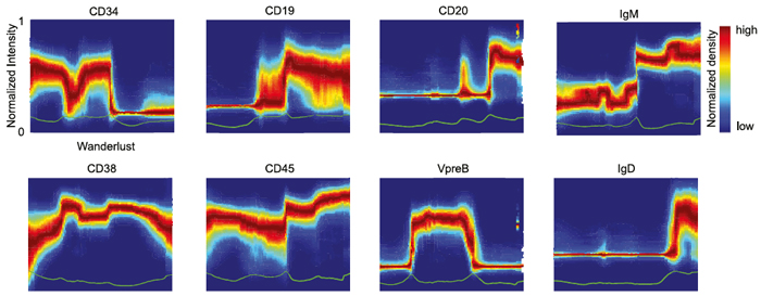
Distribution of marker expression across development

A new algorithm called Wanderlust uses single-cell measurements to detect how marker expression changes across development.

In a new paper published in the journal Cell, a team of researchers led by Dana Pe’er at Columbia University and Garry Nolan at Stanford University describes a powerful new method for mapping cellular development at the single cell level. By combining emerging technologies for studying single cells with a new, advanced computational algorithm, they have designed a novel approach for mapping development and created the most comprehensive map ever made of human B cell development. Their approach will greatly improve researchers’ ability to investigate development in cells of all types, make it possible to identify rare aberrations in development that lead to disease, and ultimately help to guide the next generation of research in regenerative medicine.

Pointing out why being able to generate these maps is an important advance, Dr. Pe’er, an associate professor in the Columbia University Department of Systems Biology and Department of Biological Sciences, explains, “There are so many diseases that result from malfunctions in the molecular programs that control the development of our cell repertoire and so many rare, yet important, regulatory cell types that we have yet to discover. We can only truly understand what goes wrong in these diseases if we have a complete map of the progression in normal development. Such maps will also act as a compass for regenerative medicine, because it’s very difficult to grow something if you don’t know how it develops in nature. For the first time, our method makes it possible to build a high-resolution map, at the single cell level, that can guide these kinds of research.”