New Technologies ×

News

Department of Systems Biology bioengineer Harris Wang describes the goals of the Human Genome Project - Write (HGP-write), an international initiative to develop new technologies for synthesizing very large genomes from scratch. 

In June 2016, a consortium of synthetic biologists, industry leaders, ethicists, and others  published a proposal in Science calling for a coordinated effort to synthesize large genomes, including a complete human genome in cell lines. The organizers of the project, called GP-write (for work in model organisms and plants) or sometimes HGP-write (for work in human cell lines), envision it as a successor to the Human Genome Project (retroactively termed HGP-read), which 25 years ago promoted rapid advances in DNA sequencing technology. As the ability to read the genome became more efficient and less expensive, it in turn enabled a revolution in how we study biology and attempt to improve human health. Now, by coordinating the development of new technologies for writing DNA on a whole-genome scale, GP-write aims to have a similarly transformative impact.

Among the paper’s authors were Virginia Cornish and Harris Wang, two members of the Columbia University Department of Systems Biology whose contributions to the field of engineering biology have in part made the idea of writing large-scale DNA sequences imaginable. We spoke with them to learn more about what GP-write hopes to accomplish, its potential benefits, and how the effort is evolving.

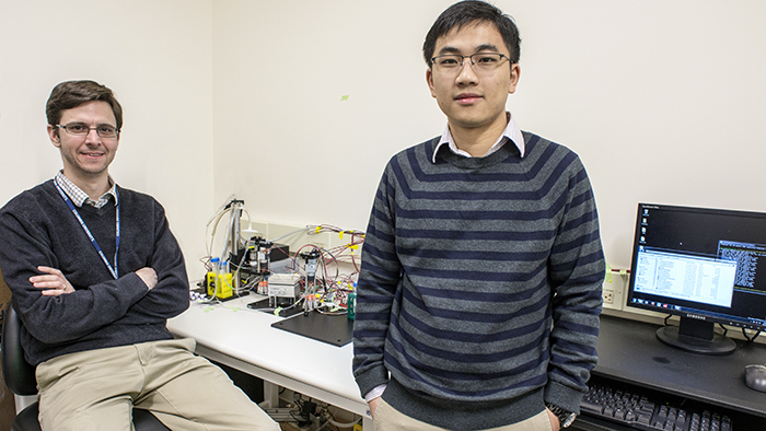
Peter Sims & Jinzhou Yuan

Assistant Professor Peter Sims and postdoctoral research scientist Jinzhou Yuan displaying their platform for automated single cell RNA-Seq. Photo: Lynn Saville.

RNA sequencing (RNA-Seq) has become a workhorse technology for research in systems biology. Unlike genome sequencing, which reveals a sample’s DNA blueprint, RNA-Seq catalogs the constantly changing transcriptome; that is, it itemizes and quantifies the complete set of messenger RNA transcripts that are present in cells at a specific time and under specific conditions. In this way, RNA-Seq makes it possible to investigate how the information encoded in the genome is functionally transformed into observable traits, and provides valuable data for defining and comparing different biological states.

Conventional RNA-Seq generates an average summary of mRNA abundance across all of the cells in a sample. Recent research, however, has created a demand for higher resolution technologies capable of generating mRNA profiles at the level of single cells. In cancer biology, for example, there is an increasingly acute awareness that gene expression in the cells that make up malignant tumors is highly heterogeneous. This suggests that in order to understand how the cells work together to drive a tumor’s cancerous behavior, scientists need better methods for characterizing the entire ecology of cells of which it is made. Being able to quantify differences in gene expression cell by cell could be one valuable way to explore such complex environments and understand how they sustain malignancy.

Although several single cell RNA-seq technologies have been unveiled in the past two years, they are expensive to operate and are not optimized to produce data on the scale that is required for systems biology research, particularly in tissue specimens with limited numbers of cells. In a new paper just published in the journal Scientific Reports, however, researchers in the laboratory of Department of Systems Biology Assistant Professor Peter Sims describe a novel approach that offers several important advantages over other existing methods.

The new, automated platform builds on previous innovations in the Sims Lab to offer a cheap, efficient, and reliable way to simultaneously measure gene expression in thousands of individual cells from a single tissue sample. Using custom designed microwell plates, microfluidics, temperature control systems, and software, the technology captures, tags, and generates a readout of the complete transcriptome in each cell, providing robust data that can then be analyzed to distinguish functional diversity among the cells in the sample. Already, the technology is playing a key role in several research projects being conducted in the Department of Systems Biology and promises to become even more powerful as the field of single cell genomics continues to evolve.

Swarmbots

In a recent paper published in Molecular Systems Biology, Kam Leong describes a two-compartment microfluidic device that consists of a chamber within which is embedded a "microbial swarmbot" that is isolated by a permeable hydrogel shell. In collaboration with Lingchong You (Duke University), Leong used the device to regulate the dynamics of a population of bacteria containing a genetically engineered switch that reacts to population size. The scale bar in panel 1 represents a length of 250 micrometers.

With a restless curiosity, Kam Leong always seems to be on the lookout for new problems to solve. A versatile biomedical engineer originally trained in chemical engineering, he has developed an impressive array of innovative nanotechnologies that have opened up new opportunities in biomedical research and drug delivery. 

The most widely known of his designs resulted from his work as a postdoc in the laboratory of MIT’s Robert Langer. While there, Leong played a critical role in the development of Gliadel, a controlled-release therapy that uses biodegradable polymer particles to deliver an anticancer drug to a brain tumor site following surgery. Since then his name has appeared on more than 70 patents covering a wide range of inventions — from microfluidics technologies, to scaffolds for growing organic tissues, to nanoscale fluorescent probes, to a method that uses nanoparticles instead of viruses for the oral delivery of gene therapies. These achievements have gained him widespread respect within the engineering community, as evidenced by his 2013 election to both the National Academy of Engineering and the National Academy of Inventors.

Dr. Leong joined Columbia University in 2014. Although his primary affiliation is with the Department of Biomedical Engineering, he was also attracted by the chance to assume an interdisciplinary faculty appointment in the Department of Systems Biology. Since his arrival he has been developing collaborations with several Systems Biology faculty members as well as other scientists at Columbia University Medical Center, and plans are underway for his lab to move into the Lasker Biomedical Research Building to better facilitate interactions with systems biology and clinical investigators. In the following interview, Leong describes why opportunities to interact with scientists in other disciplines is so important to his work, and how the kinds of technologies he has developed could be relevant for systems biology research, as well as for improving treatment of human diseases.

Cluster computer

Students participating in a new course gain experience using the Department of Systems Biology's computing cluster, a Top500 supercomputer dedicated to biological research.

As more and more biological research moves to a “big data” model, the ability to use high-performance computing platforms for analysis is rapidly becoming an essential skill set. To prepare students to work with these new tools more successfully, the Columbia University Department of Systems Biology recently partnered with the Mailman School of Public Health in launching a new graduate level class focused on providing a strong grounding in the fundamental concepts behind the technology.

Peter Sims, Sagi Shapira, and Harris Wang

Assistant Professors Peter Sims, Sagi Shapira, and Harris Wang recently moved into a new Department of Systems Biology laboratory space designed to facilitate the development of new technologies for biological and biomedical research. Photo: Lynn Saville.

The Columbia University Department of Systems Biology has opened a new experimental research hub focused on biotechnology development. Occupying one and a half floors in the Mary Woodard Lasker Biomedical Research Building at Columbia University Medical Center, the facility will promote the design and implementation of new experimental methods for the study and engineering of biological systems. It will also enable a substantial expansion of Columbia’s next-generation genome sequencing capabilities.

The first occupants of the new facility are the laboratories of Department of Systems Biology Assistant Professors Sagi Shapira, Peter Sims, and Harris Wang, along with the Genome Sequencing and Analysis Center of the JP Sulzberger Columbia Genome Center. The community is slated to grow, as currently unoccupied space will soon accommodate additional Columbia University faculty labs that are also developing new biotechnologies.

“Technology drives science,” says Department of Systems Biology Chair Andrea Califano, “and the ability to design new technologies can make it possible to answer questions that no one else can. By bringing technology-focused investigators and the Genome Center’s sequencing infrastructure together in the same physical location, our goal is that the new Lasker facility will give the Department of Systems Biology — and the entire Columbia University research community — access to unique applications for biological and biomedical research.” 

PhenoGraph

PhenoGraph, a new algorithm developed in Dana Pe'er's laboratory, proved capable of accurately identifying AML stem cells, reducing high-dimensional single cell mass cytometry data to an interpretable two-dimensional graph. Image courtesy of Dana Pe'er.

A key problem that has emerged from recent cancer research has been how to deal with the enormous heterogeneity found among the millions of cells that make up an individual tumor. Scientists now know that not all tumor cells are the same, even within an individual, and that these cells diversify into subpopulations, each of which has unique properties, or phenotypes. Of particular interest are cancer stem cells, which are typically resistant to existing cancer therapies and lead to relapse and recurrence of cancer following treatment. Finding better ways to distinguish and characterize cancer stem cells from other subpopulations of cancer cells has therefore become an important goal, for once these cells are identified, their vulnerabilities could be studied with the aim of developing better, long lasting cancer therapies.

In a paper just published online in Cell, investigators in the laboratories of Columbia University’s Dana Pe’er and Stanford University’s Garry Nolan describe a new method that takes an important step toward addressing this challenge. As Dr. Pe’er explains, “Biology has come to a point where we suddenly realize there are many more cell types than we ever imagined possible. In this paper, we have created an algorithm that can very robustly identify such subpopulations in a completely automatic and unsupervised way, based purely on high-dimensional single-cell data. This new method makes it possible to discover many new cell subpopulations that we have never seen before.”

Topology of cancer

The Columbia University Center for Topology of Cancer Evolution and Heterogeneity will combine mathematical approaches from topological data analysis with new single-cell experimental technologies to study cellular diversity in solid tumors. Image courtesy of Raul Rabadan.

The National Cancer Institute’s Physical Sciences in Oncology program has announced the creation of a new center for research and education based at Columbia University. The Center for Topology of Cancer Evolution and Heterogeneity will develop and utilize innovative mathematical and experimental techniques to explore how genetic diversity emerges in the cells that make up solid tumors. In this way it will address a key challenge facing cancer research in the age of precision medicine — how to identify the clonal variants within a tumor that are responsible for its growth, spread, and resistance to therapy. Ultimately, the strategies the Center develops could be used to identify more effective biomarkers of disease and new therapeutic strategies.

Fluidigm C1 Single-Cell Plate

At the core of the Fluidigm C1 Single-Cell Auto Prep System is a 96-well plate containing microfluidics. After individual cells are isolated in their own wells, the device amplifies their cDNA for genome-wide gene expression profiling. Scientists at the Columbia Genome Center are developing methods for addressing the technical and analytical challenges of single-cell RNA sequencing, and have begun generating some exciting data.

Since the invention of the first microscope, a procession of new technologies has enabled scientists to study individual cells at increasingly fine levels of detail. The last two years have witnessed an important next stage in this evolution, with the arrival of the first devices for genetically profiling single cells on a genome-wide scale.

The first commercial product in this field is the Fluidigm C1 Single-Cell Auto Prep System, which uses microfluidics to isolate single cells and offers the ability to generate gene expression profiles for up to 96 cells at a time. But because of the novelty of the technology and the inherent difficulties of working with single cells, it has presented a number of technical challenges for researchers interested in exploring biology at this level.

Now, scientists at the JP Sulzberger Columbia Genome Center led by Assistant Professors Peter Sims and Yufeng Shen have developed an experimental and computational pipeline that optimizes the C1’s capabilities. And even as they work to solve some of the challenges that are inherent to single-cell research, their approach has begun generating some exciting data for studying genetics in a variety of cell types.

Dana Pe'er and Kyle Allison

Dana Pe'er has received the Pioneer Award for high-risk, high-reward research, and postdoctoral scientist Kyle Allison has won an Early Independence Award.

Two members of the Columbia University Department of Systems Biology have been named recipients of NIH Director’s Awards from the National Institutes of Health Common Fund.

Associate Professor Dana Pe’er is one of 10 winners of the 2014 NIH Director’s Pioneer Awards. The Pioneer Awards provide up to $2.5 million over 5 years to support exceptionally creative investigators who are pursuing “high risk, high reward” science that holds great potential to transform biomedical or behavioral research. The award will support an ambitious new project to develop the technological and computational methods necessary to create a comprehensive, high-resolution atlas of development for all cell types in the human body.

In addition, Kyle Allison, a postdoctoral scientist in the laboratory of Professor Saeed Tavazoie, has received the NIH Director’s Early Independence Award. (Dr. Tavazoie is also a past winner of the Pioneer Award.) This program enables outstanding young investigators who have recently completed their PhD’s to move rapidly into independent research positions. Dr. Allison is one of just 17 scientists to receive this award this year. In combination with the Department of Systems Biology Fellows program, this five-year, $1.25 million grant will allow him to open his own laboratory at Columbia and pursue independent research to investigate the problem of bacterial persistence. He is the second Department of Systems Biology investigator to receive the Early Independence Award, joining Assistant Professor Harris Wang in being recognized with this honor.

“Having four recipients of NIH Director’s Awards within the Department of Systems Biology — and particularly two in one year — is quite remarkable,” said Department Chair Andrea Califano. “I think it’s a testimony to the timeliness of the perspectives and tools that systems biology offers and to the high quality of research being conducted at Columbia. I look forward to the discoveries that will undoubtedly come from Dana’s and Kyle’s extremely exciting efforts.”

Harris Wang

As a graduate student in George Church’s lab at Harvard University, Harris Wang developed MAGE, a revolutionary tool for the field of synthetic biology that made it possible to introduce genomic mutations into E. coli cells in a highly specific and targeted way. Now an Assistant Professor in the Columbia University Department of Systems Biology, Dr. Wang recently published a paper in ACS Synthetic Biology that introduces an important advance in the MAGE technology. The new technique, called (MO)-MAGE, uses microarrays to engineer pools of oligonucleotides that, once amplified and integrated into a genome, can generate thousands or even millions of highly controlled mutations simultaneously. This new method offers a cost-effective way for designing and producing large numbers of genomic variants and provides an efficient platform for experimentally exploring genome-wide landscapes of mutations in bacteria and optimizing the organisms’ biochemical capabilities.

In the following interview, Dr. Wang explains the origins of the new technology, and discusses what he sees as the remarkable potential it holds for both basic biological research and industrial applications of synthetic biology.

How are MAGE and (MO)-MAGE different from more traditional methods in genome engineering?

In traditional genome engineering, researchers would induce genome perturbations randomly. For example, you might use ultraviolet radiation or a mutagen to generate mutations and then do a selection experiment to compare and isolate cells with different genotypes based on how they respond to specific stimuli. The problem with this approach, though, is that you have no way to control what mutations occur, even if you know the mutation you are interested in investigating.

(MO)-MAGE offers a cost-effective and efficient way to simultaneously mutate large numbers of genes in a targeted way.

geWorkbench screenshot

A new version of geWorkbench lets researchers access a range of powerful, integrated bioinformatics tools using a standard web browser. Here, an ARACNe-generated gene regulatory network is displayed using the Cytoscape Web plugin.

Since its creation in 2005, investigators in Columbia University’s Center for the Multiscale Analysis of Genomic and Cellular Networks (MAGNet) have developed a large number of computational tools for studying biological systems from the perspectives of structural biology and systems biology. To consolidate and disseminate these tools to the wider research community, MAGNet developed geWorkbench (genomics Workbench), a free, open-source bioinformatics application that gathers all of the Center’s software and databases into one integrated software platform. These include applications for the analysis of cellular regulatory networks, protein structure, DNA and protein sequences, gene expression, and other kinds of biological data.

Initially, geWorkbench was made available as a software package that users could install and run on their local computers. Now, in a major upgrade, MAGNet has released a web-based version that makes these tools accessible through a browser interface.

Distribution of marker expression across development

A new algorithm called Wanderlust uses single-cell measurements to detect how marker expression changes across development.

In a new paper published in the journal Cell, a team of researchers led by Dana Pe’er at Columbia University and Garry Nolan at Stanford University describes a powerful new method for mapping cellular development at the single cell level. By combining emerging technologies for studying single cells with a new, advanced computational algorithm, they have designed a novel approach for mapping development and created the most comprehensive map ever made of human B cell development. Their approach will greatly improve researchers’ ability to investigate development in cells of all types, make it possible to identify rare aberrations in development that lead to disease, and ultimately help to guide the next generation of research in regenerative medicine.

Pointing out why being able to generate these maps is an important advance, Dr. Pe’er, an associate professor in the Columbia University Department of Systems Biology and Department of Biological Sciences, explains, “There are so many diseases that result from malfunctions in the molecular programs that control the development of our cell repertoire and so many rare, yet important, regulatory cell types that we have yet to discover. We can only truly understand what goes wrong in these diseases if we have a complete map of the progression in normal development. Such maps will also act as a compass for regenerative medicine, because it’s very difficult to grow something if you don’t know how it develops in nature. For the first time, our method makes it possible to build a high-resolution map, at the single cell level, that can guide these kinds of research.”

Saeed Tavazoie

One of the defining features of systems biology has been its integration of computational and experimental methods for probing networks of molecular interactions. The research of Saeed Tavazoie, a professor in the Columbia University Department of Systems Biology, has been emblematic of this approach. After undergraduate studies in physics, he became fascinated by the processes that govern gene expression, particularly in understanding how gene expression is regulated by information encoded in the genome. Since then, his multidisciplinary approach to research has generated important insights into the principles that orchestrate genome regulation, as well as a number of novel algorithms and technologies for exploring this complex landscape.

In this conversation, Dr. Tavazoie discusses his research in the areas of gene transcription, post-transcriptional regulation, and molecular evolution, as well as some innovative technologies and experimental methods his lab has developed.

Peter SimsPeter Sims, an assistant professor in the Columbia University Department of Systems Biology, has been named Associate Director for Novel Technologies at the JP Sulzberger Columbia Genome Center. In this role he will devise, direct, and implement strategies for incorporating new high-throughput experimental methods into the research done at the Genome Center.

Trained as a physical chemist, Dr. Sims has been developing a number of innovative technologies for studying single cells in a high-throughput setting. Using a type of microfluidics called soft lithography, his laboratory has designed a method for creating arrays composed of wells just tens of microns in diameter, small enough to isolate and perform high-throughput experiments on individual cells.  

Appointing Dr. Sims to his new role will enable the Columbia Genome Center to develop a variety of new applications that will benefit researchers across the Columbia University community. 

Imaging synapses

In a test of the Columbia Genome Center's high-content microscopy system, computational image analysis confirmed a high degree of colocalization of green fluorescent protein-labeled synapsin and the dye FM4-64. The researchers plan a high-throughput screen to identify fluroescent small molecules capable of targeting synapses.

Synapses mediate communication between neurons in the brain, making them critical components for neurological activity. Research has shown that synaptic loss and dysfunction play roles in a number of debilitating brain disorders — including Alzheimer’s disease, major depressive disorder, and autism — but currently no effective method exists for identifying and imaging individual synapses in living human brains. Being able to locate and quantify synapses in patients could greatly improve the diagnosis and monitoring of disease, and potentially offer new approaches for treatment.

Clarissa Waites, an assistant professor of pathology and cell biology at the Columbia University College of Physicians & Surgeons, and Dalibor Sames in the Columbia University Department of Chemistry, have recently embarked on a collaboration with the Columbia Genome Center High-Throughput Screening Facility with the goal of identifying small fluorescent molecules that can selectively localize to synapses. If successful, this project could for the first time provide a method for targeting and imaging synapses in the living human brain.

viSNE

viSNE reveals the progression of cancer in a sample of cells taken from a patient with acute myeloid leukemia. Cells are colored according to intensity of expression of the indicated cell markers, enabling the comparison of expression patterns before and after relapse. For example, Fit3 is expressed primarily in the diagnosis sample, while CD34 emerges in the relapse sample.

Researchers in the Columbia Initiative in Systems Biology have developed a computational method that enables scientists to visualize and interpret high-dimensional data produced by single-cell measurement technologies such as mass cytometry. The method, called viSNE (visual interactive Stochastic Neighbor Embedding), has just been published in the online edition of Nature Biotechnology. It has particular relevance to cancer research and therapeutics. As Columbia University Medical Center reports:

Researchers now understand that cancer within an individual can harbor subpopulations of cells with different molecular characteristics. Groups of cells may behave differently from one another, including in how they respond to treatment. The ability to study single cells, as well as to identify and characterize subpopulations of cancerous cells within an individual, could lead to more precise methods of diagnosis and treatment.

“Our method not only will allow scientists to explore the heterogeneity of cancer cells and to characterize drug-resistant cancer cells, but also will allow physicians to track tumor progression, identify drug-resistant cancer cells, and detect minute quantities of cancer cells that increase the risk of relapse,” said co-senior author Dana Pe’er, associate professor of biological sciences and systems biology at Columbia.