Next-Generation Sequencing ×

News

Raul Rabadan
Raul Rabadan

Systems Biology Professor Raul Rabadan, Phd , has been awarded a Philip A. Sharp Innovation in Collaboration award from Stand Up to Cancer (SU2C) , a group established by film and media leaders to fund cancer research projects that have the potential to quickly deliver new therapies to patients. Dr. Rabadan has received the award jointly with collaborator Dan A. Landau, MD, PhD, of Weill Cornell Medicine.

A theoretical physicist whose expertise lies in the cross section of mathematical genomics, tumor evolution, and cancer research, Dr. Rabadan will work together with Dr. Landau on their winning project, “Cupid-seq—high throughput transcriptomic spatial mapping of immune-tumor interactions in the micro-environment.”  The investigators will devise a novel sequencing technique and computational method for better understanding immune recognition mechanism in glioblastoma. Dr. Rabadan is currently a principal investigator on the SU2C-National Science Foundation Drug Combination Convergence Team and Dr. Lau is a 2016 recipient of a SU2C Innovative Research Grant.

The idea behind the Sharp Awards is to fund projects that involved SU2C researchers who have not yet worked together, including collaborations between members of Dream Teams, Research Teams, and Convergence teams, and between SU2C members and recipients of its innovative research grants. The latter are typically early-career investigators. The Sharp Awards are named after Sharp, Nobel laureate and institute professor at MIT and the Koch Institute for Integrated Cancer Research, to honor the emphasis he has placed on the importance of collaborative research. 

Dr. Rabadan, who also is professor of biomedical informatics at Columbia University, is one of 10 recipients of this year’s Sharp Awards; investigators hail from the U.S., Canada, and the Netherlands, and projects are being funded from a pool of $1.25 million. 

Coauthors
Study lead coauthors Nathan Johns (left), systems biology graduate student in the Wang Lab, and Antonio Gomes, former member of the Wang Lab, now at Memorial Sloan Kettering Cancer Center.

Advances in synthetic biology have already spurred innovation in the areas of drug development, chemical production and health diagnostics. To help push the field even further, and potentially at a more rapid pace, a new, comprehensive resource devised by Columbia University investigators will help synthetic biologists better engineer designs for complex biological systems.

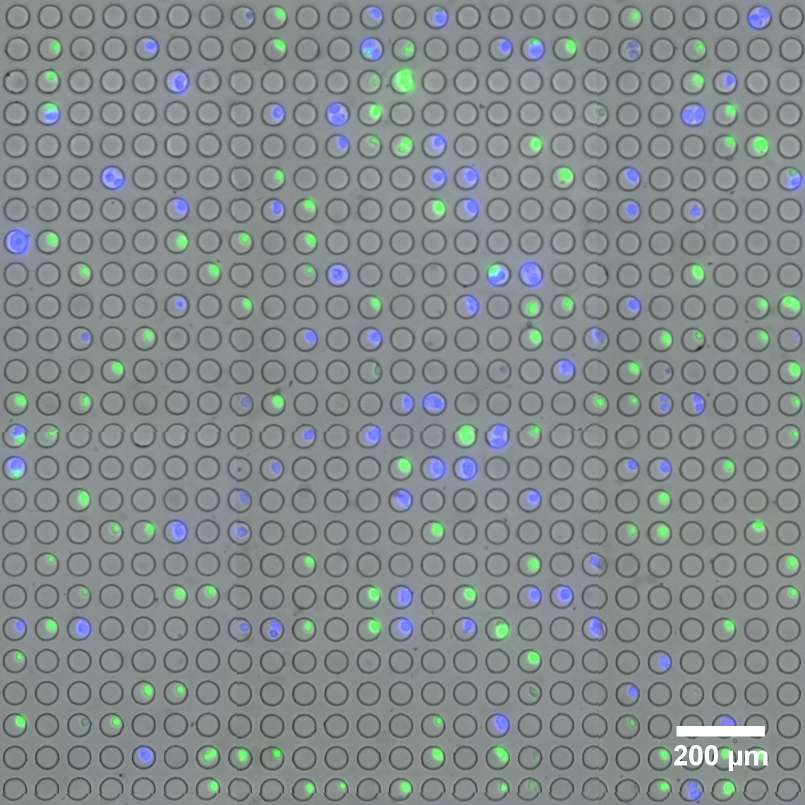
A team of researchers, led by Harris Wang, PhD, assistant professor of systems biology and of pathology and cell biology, report the characterization and analysis of thousands of bacterial regulatory elements in different species of bacteria. The paper , published March 19, appears in Nature Methods .

Synthetic biology employs well-characterized genetic parts to assemble gene circuits with specific functions, such as producing chemicals or sensing the environment. The toolbox of genetic parts to make functioning genetic circuits, however, has been limiting. A key shortfall is the availability of precisely measured regulatory sequences-segments of DNA responsible for dialing up or dialing down the expression of proteins within an organism. For many commercially useful bacteria, tuning gene expression has been challenging because of a lack of reliable regulatory sequences. 

"Synthetic biology is now at a precipice where we are not just demonstrating proof-of-concept in the laboratory but we're moving toward real-world applications," says Nathan Johns , lead coauthor of the paper, a member of the Wang Lab and a graduate student in the Department of Systems Biology at Columbia. "To facilitate this, having a wide array of useful genetic components and measurement techniques-in our case, regulatory sequences-are extremely helpful." 

New NIH Grant to Accelerate Commercialization of Single-Cell Analysis Platform

fluorescently-labeled live cells
Microwell array flow cell device loaded with fluorescently-labeled live cells. (Image provided by the Sims Lab.)

The field of single-cell RNA sequencing is moving at a fast clip. Adding to its rapid advance is a novel platform for linking optical imaging with high-throughput single-cell sequencing devised by researchers in the Sims Lab at Columbia’s Department of Systems Biology.

The new technology, developed by Peter Sims, PhD , assistant professor of systems biology, and postdoctoral research scientist, Jinzhou Yuan, PhD , enables live cell imaging and RNA sequencing simultaneously of the same individual cell on a large scale and at low cost. Jointly awarded a $1.5 million grant funded by the National Institutes of Health’s SBIR program, the Sims Lab and Cell Microsystems are collaborating to build and test the device, with the goal of bringing to market a fully-integrated system capable of imaging thousands of single cells and preparing them for genomic analysis.

The proposed system will integrate Cell Microsystems’ proprietary CellRaft Technology with the Sims Lab’s novel approaches to tracking single cells with so-called optical barcodes.

In single-cell RNA sequencing, “State-of-the-art technology now allows us to routinely process thousands of individual cells and obtain their genome-wide mRNA expression profiles,” notes Dr. Yuan. “However, cells that share similar expression profiles at the mRNA level may be distinct from each other based on features obtainable by optical microscopy, such as morphology, motility, and fluorescent labels. A more comprehensive description of each cell obtained by both imaging and sequencing may help us further refine cell type and state.”

HTS
Research scientist Hai Li holds up a 384-well plate, pictured in front of Columbia Genome Center's Hamilton Star automation system for HTS; Image credit: Systems Biology

Drug screening and analysis is critical in advancing research and discovery of cancer therapeutics. To this end, a Systems Biology-led team of investigators has recently developed PLATE-Seq, a new technique for low-cost, bulk mRNA sequencing. Coupled with genome-wide regulatory network analysis, the novel PLATE-Seq method advances the goal of providing cancer patients with personalized treatment.

Developed by the labs of Peter Sims and Andrea Califano , PLATE-Seq stands for “pooled library amplification for transcriptome expression” sequencing, and enables genome-wide mRNA profiling specifically designed to complement high-throughput screening assays. High-throughput screening, or HTS, represents a key component of drug discovery and technology used widely in biomedical research. Due to cost or complexity, most screens are still performed using low-complexity reporters, such as cell viability, protein-protein interactions and cell growth, for example, but there is a growing need to couple this screening protocol with genome-wide reporters, to measure the activity of many proteins across the genome.

“Our PLATE-Seq method helps us generate a more comprehensive portrait of drug activity,” said Dr. Sims, assistant professor of systems biology, known for his innovative work in single-cell RNA sequencing. “We’ve been able to show that our technique cuts the cost for gene expression profiling considerably, by incorporating a method we devised for ‘barcoding’ cDNA samples and combining this with computational methods from the Califano Lab that are highly effective on low-coverage sequencing data. This method allows us to sequence 96 samples per plate and 768 samples per sequencing run.”

On average, PLATE-Seq reduces the cost of genome-wide screening from around $400 per sample down to approximately $25 per sample. Genome-wide sequencing is important in advancing our understanding and prediction of disease and in identifying potential treatments.

Peter Sims Lab Wins CZI Award

Assistant Professor Peter Sims and postdoctoral research scientist Jinzhou Yuan displaying their platform for automated single-cell RNA sequencing. (Photo: Lynn Saville)

Assistant Professor Peter Sims, PhD , has been awarded an inaugural Chan Zuckerberg Initiative (CZI) award for gene sequencing research that will help advance the Human Cell Atlas project. Launched in 2016 by a cohort of world-leading scientists, the Human Cell Atlas is a high-profile endeavor whose goal is to identify and define every cell type of the human body and create a collection of maps that will describe the cellular basis of health and disease.

With the support of CZI, founded by Facebook CEO Mark Zuckerberg and his wife, Priscilla Chan, Dr. Sims and his group in the Department of Systems Biology will receive grant funding to pilot a revolutionary technique for high throughput single-cell sequencing. Called SCOPE-Seq, the novel, economical method conducts RNA sequencing coupled with live imaging of the same individual cell on a large scale.

“We hope that our approach will provide functional insights into the novel cell types that will be discovered by the Human Cell Atlas effort that cannot be obtained from genomic analysis alone,” said Dr. Sims.

Raul Rabadan
Raul Rabadan

Fellow Systems Biology Professor Raul Rabadan, PhD , who directs the Center for Topology of Cancer Evolution and Heterogeneity, also is gaining support from CZI in a collaboration led by Tom Maniatis, PhD, who won the grant for their research to construct an atlas of gene activity of all cells in the human spinal cord. Dr. Maniatis chairs the Department of Biochemistry and Molecular Biophysics, directs Columbia's Precision Medicine Initiative and is a principal investigator at Columbia's Mortimer B. Zuckerman Mind Brain Behavior Institute.

Staphylococcus epidermis
Interactions between human cells and the bacteria that inhabit our bodies can affect health. Here, Staphylococcus epidermis binds to nasal epithelial cells. (Image courtesy of Sheetal Trivedi and Sean Sullivan.)

Launched in 2014 by investigators in the Mailman School of Public Health, the CUMC Microbiome Working Group brings together basic, clinical, and population scientists interested in understanding how the human microbiome—the ecosystems of bacteria that inhabit and interact with our tissues and organs—affects our health. Computational biologists in the Department of Systems Biology have become increasingly involved in this interdepartmental community, contributing expertise in analytical approaches that make it possible to make sense of the large data sets that microbiome studies generate.

Deep sequencing class

A new team-taught course covers both the experimental and analytical basics of next-generation sequencing. Assistant Professor Chaolin Zhang led the discussion in a recent class. Photo: Lynn Saville.

As the cost of next-generation sequencing has fallen, it has become a ubiquitous and indispensable tool for research across the biomedical sciences. DNA and RNA sequencing — along with other technologies for profiling phenomena such as de novo mutations, protein-nucleic acid interactions, chromatin accessibility, ribosome activity, and microRNA abundance — now make it possible to observe multiple layers of cellular function on a genome-wide scale.

Regardless of a biologist’s chosen area of investigation, such methods have made it possible to explore many exciting new kinds of problems. At the same time, however, it has also dramatically transformed the expertise that young scientists need to develop in order to participate in cutting-edge biological research. Bringing students up to speed with the pace of change in next-generation sequencing has posed a particular challenge for educators.

Now, a new multidisciplinary, graduate-level course organized by the Columbia University Department of Systems Biology is enabling young investigators to begin incorporating these powerful new tools into their studies and future research. Designed by assistant professors Yufeng Shen, Peter Sims, and Chaolin Zhang, the course covers both the experimental principles of next-generation sequencing and key statistical methods for analyzing the enormous datasets that such technologies produce. In this way, it gives students a strong grounding in principles that are critical for more advanced graduate courses as well as the ability to begin applying deep sequencing technologies to investigate the questions they are interested in pursuing.

As Dr. Sims explains, “Whether you are a graduate student in systems biology, biochemistry, or microbiology, the chance that you are going to be doing next-generation sequencing is pretty high. At the same time, it’s completely not taught at the undergraduate level. There is no text book nor is there any time in a typical undergraduate biology curriculum to get into this in any kind of detail. Even at top-tier universities students come into graduate school without having any experience with it, and often they’re expected to jump right into this kind of research. We decided that this was a problem we had to fix.”

Alex Lachmann
Alex Lachmann during his presentation to the RNA-Seq "boot camp."

In June 2015, the Columbia University Department of Systems Biology held a five-part lecture series focusing on advanced applications of RNA-Seq in biological research. The talks covered topics such as the use of RNA-Seq for studying heterogeneity among single cells, RNA-Seq experimental design, statistical approaches for analyzing RNA-Seq data, and the utilization of RNA-Seq for the prediction of molecular interaction networks. The speakers and organizers have compiled a list of lecture notes and study materials for those wishing to learn more. Click on the links below for more information.

Tracking clones

After identifying T cell clones that react against donated kidney tissue in vitro, new computational methods developed in Yufeng Shen's Lab are used to track their frequency following organ transplant. The findings can help to predict transplant rejection or tolerance.

When a patient receives a kidney transplant, a battle often ensues. In many cases, the recipient’s immune system identifies the transplanted kidney as a foreign invader and mounts an aggressive T cell response to eliminate it, leading to a variety of destructive side effects. To minimize complications, many transplant recipients receive drugs that suppress the immune response. These have their own consequences, however, as they can lead to increased risk of infections. For these reasons, scientists have been working to gain a better understanding of the biological mechanisms that determine transplant tolerance and rejection. This knowledge could potentially improve physicians’ ability to predict the viability of an organ transplant and to provide the best approach to immunosuppression therapy based on individual patients’ immune system profiles.

Yufeng Shen, an assistant professor in the Columbia University Department of Systems Biology and JP Sulzberger Columbia Genome Center, together with Megan Sykes, director of the Columbia Center for Translational Immunology at the Columbia University College of Physicians and Surgeons, recently took an encouraging step toward this goal. In a paper published in Science Translational Medicine, they report that the deletion of specific donor-resistant T cell clones in the transplant recipient can support tolerance of a new kidney. Critical to this discovery was the development of a new computational genomics approach by the Shen Lab, which makes it possible to track how frequently rare T cell clones develop and how their frequencies change following transplantation. The paper suggests both a general strategy for understanding the causes of transplant rejection and a means of identifying biomarkers for predicting how well a transplant recipient will tolerate a new kidney.

ALK-negative ALCL mutation map
A map of mutations observed in ALK-negative anaplastic large cell lymphoma. (Credit: Dr. Rabadan)

The following article is reposted with permission from the Columbia University Medical Center Newsroom. Find the original here.

The first-ever systematic study of the genomes of patients with ALK-negative anaplastic large cell lymphoma (ALCL), a particularly aggressive form of non-Hodgkin’s lymphoma (NHL), shows that many cases of the disease are driven by alterations in the JAK/STAT3 cell signaling pathway. The study also demonstrates, in mice implanted with human-derived ALCL tumors, that the disease can be inhibited by compounds that target this pathway, raising hopes that more effective treatments might soon be developed. The study, led by researchers at Columbia University Medical Center (CUMC) and Weill Cornell Medical College, was published today in the online edition of Cancer Cell.

Gut bacteria

Photo by David Gregory and Debbie Marshall, Wellcome Images. 

Recent deep sequencing studies are providing an increasingly detailed picture of the genetic composition of the human microbiome, the diverse collection of bacterial species that inhabit the gut. At the same time, however, little is known about the dynamics of these colonies, particularly why certain microbial strains outcompete others in the same environment. In a new paper published in the journal Molecular Systems Biology, Department of Systems Biology Assistant Professor Harris Wang, in collaboration with Georg Gerber and researchers at Harvard University, report on their development of the first method for using functional metagenomics to identify genes within commensal bacterial genomes that give them an evolutionary fitness advantage.

Sequence of genomic alterations in CLLA graph representing the sequence of genomic alterations in chronic lymphocytic leukemia (CLL). Each node represents a mutation, with arrows indicating temporal relationships between them. The size of the nodes indicates the number of patients in the study who exhibited the alteration, while the thickness of the lines shows how often the temporal relationships between nodes were seen. The method the researchers use enabled them to identify multiple, distinct evolutionary patterns in CLL.

As biologists have gained a better understanding of cancer, it has become clear that tumors are often driven not by a single mutation, but by a series of genetic changes that correspond to particular stages of cancer progression. In this sense, a tumor is constantly evolving, with different groups of cells that harbor distinctive mutations multiplying at different rates, depending on their fitness for particular disease states. As the search for more effective cancer diagnostics and therapies continues, one key question is how to disentangle the order in which mutations occur in order to understand how tumors change over time. Being able to predict how a tumor will behave based on signs seen early in the course of disease could enable the development of new diagnostics that could better inform treatment planning.

In a paper just published in the journal eLife, a team of investigators led by Department of Systems Biology Associate Professor Raul Rabadan reports on a new computational strategy for addressing this challenge. Their framework, called tumor evolutionary directed graphs (TEDG), considers next-generation sequencing data from tumor samples from a large number of patients. Using TEDG to analyze cancer cells in patients with chronic lymphocytic leukemia (CLL), they were able to develop a model of how the disease’s mutational landscape changes from its initial onset to its late stages. Their findings suggest that CLL may not be just the result of a single evolutionary path, but can evolve in alternative ways.

Fluidigm C1 Single-Cell Plate

At the core of the Fluidigm C1 Single-Cell Auto Prep System is a 96-well plate containing microfluidics. After individual cells are isolated in their own wells, the device amplifies their cDNA for genome-wide gene expression profiling. Scientists at the Columbia Genome Center are developing methods for addressing the technical and analytical challenges of single-cell RNA sequencing, and have begun generating some exciting data.

Since the invention of the first microscope, a procession of new technologies has enabled scientists to study individual cells at increasingly fine levels of detail. The last two years have witnessed an important next stage in this evolution, with the arrival of the first devices for genetically profiling single cells on a genome-wide scale.

The first commercial product in this field is the Fluidigm C1 Single-Cell Auto Prep System, which uses microfluidics to isolate single cells and offers the ability to generate gene expression profiles for up to 96 cells at a time. But because of the novelty of the technology and the inherent difficulties of working with single cells, it has presented a number of technical challenges for researchers interested in exploring biology at this level.

Now, scientists at the JP Sulzberger Columbia Genome Center led by Assistant Professors Peter Sims and Yufeng Shen have developed an experimental and computational pipeline that optimizes the C1’s capabilities. And even as they work to solve some of the challenges that are inherent to single-cell research, their approach has begun generating some exciting data for studying genetics in a variety of cell types.

Ashkenazi Population Bottleneck Model
The consortium’s model of Ashkenazi Jewish ancestry suggests that the population’s history was shaped by three critical bottleneck events. The ancestors of both populations underwent a bottleneck sometime between 85,000 and 91,000 years ago, which was likely coincident with an Out-of-Africa event. The founding European population underwent a bottleneck at approximately 21,000 years ago, beginning a period of interbreeding between individuals of European and Middle Eastern ancestry. A severe bottleneck occurred in the Middle Ages, reducing the population to under 350 individuals. The modern-day Ashkenazi community emerged from this group.

An international research consortium led by Associate Professor Itsik Pe’er has produced a new panel of reference genomes that will significantly improve the study of genetic variation in Ashkenazi Jews. Using deep sequencing to analyze the genomes of 128 healthy individuals of Ashkenazi Jewish origin, The Ashkenazi Genome Consortium (TAGC) has just published a resource that will be much more effective than previously available European reference genomes for identifying disease-causing mutations within this historically isolated population. Their study also provides novel insights into the historical origins and ancestry of the Ashkenazi community. A paper describing their study has just been published online in Nature Communications.

The dataset produced by the consortium provides a high-resolution baseline genomic profile of the Ashkenazi Jewish population, which they revealed to be significantly different from that found in non-Jewish Europeans. In the past, clinicians’ only option for identifying disease-causing mutations in Ashkenazi individuals was to compare their genomes to more heterogeneous European reference sets. This new resource accounts for the historical isolation of this population, and so will make genetic screening much more accurate in identifying disease-causing mutations.

In an article that appears on the website of Columbia University’s Fu Foundation School of Engineering and Computer Science, Dr. Pe’er explains:

“Our study is the first full DNA sequence dataset available for Ashkenazi Jewish genomes... With this comprehensive catalog of mutations present in the Ashkenazi Jewish population, we will be able to more effectively map disease genes onto the genome and thus gain a better understanding of common disorders. We see this study serving as a vehicle for personalized medicine and a model for researchers working with other populations.”

In addition to offering an important resource for such future translational and clinical research, the paper’s findings also provide new insights that have implications for the much debated question of how European and Ashkenazi Jewish populations emerged historically.

Illumina NextSeq 500 at Columbia University

As genome sequencing technologies evolve, the JP Sulzberger Columbia Genome Center continues to provide the Columbia University biological and biomedical research community access to state-of-the-art tools. In its most recent acquisition, the Genome Center has just installed two Illumina NextSeq 500 sequencers. The NextSeq 500 is a flexible and efficient desktop sequencer that offers powerful high-throughput sequencing capabilities.

Columbia investigators who are experienced with the Illumina next-generation sequencing platform can now schedule time to use the NextSeq 500 for their own research. 

Tuuli LappalainenTuuli Lappalainen has joined Columbia University as an assistant professor in the Department of Systems Biology. Dr. Lappalainen is a specialist in the analysis of RNA sequencing data, with research interests including functional variation in the human genome, population genetic background of variation in the human genome, and interpretation of genome function.

Dr. Lappalainen joins the Department of Systems Biology in co-appointment with the New York Genome Center (NYGC), where she will also serve as a Junior Investigator and Core Member. Based in lower Manhattan, NYGC is a consortium made up primarily of New York-area institutions that is designed to translate promising genomics-based research into new strategies for treating, preventing, and managing disease. This co-appointment with Columbia University — an institutional founding member of the NYGC — will enhance collaboration between the two institutions. (Read an interview with Dr. Lappalainen at the New York Genome Center website.)

Dr. Lappalainen earned her PhD in genetics at the University of Helsinki, Finland, and held appointments as a postdoctoral researcher in at the University of Geneva Medical School, Switzerland and at the Stanford University School of Medicine. She is the chair of the analysis group for the Genetic European Variation in Health and Disease (Geuvadis) Consortium’s RNA sequencing project, a member of the analysis group for the National Institute of Health’s Genotype Tissue Expression (GTEx) project, and a member of the analysis and functional interpretation groups for the 1000 Genomes Project.

Rabadan, Nature Genetics

An analysis of all gene mutations in nearly 140 brain tumors has uncovered most of the genes responsible for driving glioblastoma. The analysis found 18 new driver genes (labeled red), never before implicated in glioblastoma and correctly identified the 15 previously known driver genes (labeled blue). The graphs show mutated genes that are commonly found in varying numbers in glioblastoma (left), that frequently contain insertions (middle), and that frequently contain deletions (right). Genes represented by blue dots in the graphs were statistically most likely to be driver genes.

A team of Columbia University Medical Center researchers has identified 18 new genes responsible for driving glioblastoma multiforme, the most common—and most aggressive—form of brain cancer in adults. The study was published August 5, 2013, in the journal Nature Genetics.

The Columbia team used a combination of high-throughput DNA sequencing and a new method of statistical analysis developed by co-author Raul Rabadan, an assistant professor in the Department of Systems Biology, to generate a short list of candidate gene mutations that were highly likely to drive cancer, as opposed to mutations that have no effect.

Considering these results along with a previous study this group conducted, Rabadan and collaborators Antonio Iavarone and Anna Lasorella point out that approximately 15% of glioblastomas could now be targeted with drugs that have already been approved by the FDA. As Lasorella remarks in an article for the CUMC Newsroom, “There is no reason why these patients couldn’t receive these drugs now in clinical trials.”