Cancer ×

News

Project GENIE
Richard Carvajal (left) and Raul Rabadan are lead PIs on Project GENIE

Columbia University Irving Medical Center (CUIMC) has recently joined 11 new institutions to collaborate on Project GENIE, an ambitious consortium organized by the  American Association for Cancer Research . An international cancer registry built through data sharing,  Project GENIE , which stands for Genomics Evidence Neoplasia Information Exchange, brings together leading institutions in cancer research and treatment in order to provide the statistical power needed to improve clinical decision-making, particularly in the case of rare cancers and rare genetic variants in common cancers. Additionally, the registry, established in 2016, is powering novel clinical and translational research. In its first two years, Project GENIE has been able to accumulate and make public more than 39,000 cancer genomic records, de-identified to maintain patient privacy. 

From Code to Cure

Columbia Magazine

Published Spring 2018 cover story , Columbia Magazine

As reported by David J. Craig, senior editor at Columbia Magazine , we are living in the age of big data, and with every link we click, every message we send, and every movement we make, we generate torrents of information. In the past two years, the world has produced more than 90 percent of all the digital data that has ever been created. New technologies churn out an estimated 2.5 quintillion bytes per day. 

Today, researchers at Columbia University Irving Medical Center (CUIMC) are using the power of data to identify previously unrecognized drug side effects; they are predicting outbreaks of infectious diseases by monitoring Google search queries and social-media activity; and they are developing novel cancer treatments by using predictive analytics to model the internal dynamics of diseased cells. These ambitious projects, many of which involve large interdisciplinary teams of computer scientists, engineers, statisticians, and physicians, represent the future of academic research.

Craig covers Dr. Nicholas Tatonetti's work involving prescription drug safety and his innovative use of digital health and clinical records and Dr. Andrea Califano's unconventional computational approaches in advancing cancer research.

To read the full article , visit the online issue of Columbia Magazine

Raul Rabadan
Raul Rabadan

Systems Biology Professor Raul Rabadan, Phd , has been awarded a Philip A. Sharp Innovation in Collaboration award from Stand Up to Cancer (SU2C) , a group established by film and media leaders to fund cancer research projects that have the potential to quickly deliver new therapies to patients. Dr. Rabadan has received the award jointly with collaborator Dan A. Landau, MD, PhD, of Weill Cornell Medicine.

A theoretical physicist whose expertise lies in the cross section of mathematical genomics, tumor evolution, and cancer research, Dr. Rabadan will work together with Dr. Landau on their winning project, “Cupid-seq—high throughput transcriptomic spatial mapping of immune-tumor interactions in the micro-environment.”  The investigators will devise a novel sequencing technique and computational method for better understanding immune recognition mechanism in glioblastoma. Dr. Rabadan is currently a principal investigator on the SU2C-National Science Foundation Drug Combination Convergence Team and Dr. Lau is a 2016 recipient of a SU2C Innovative Research Grant.

The idea behind the Sharp Awards is to fund projects that involved SU2C researchers who have not yet worked together, including collaborations between members of Dream Teams, Research Teams, and Convergence teams, and between SU2C members and recipients of its innovative research grants. The latter are typically early-career investigators. The Sharp Awards are named after Sharp, Nobel laureate and institute professor at MIT and the Koch Institute for Integrated Cancer Research, to honor the emphasis he has placed on the importance of collaborative research. 

Dr. Rabadan, who also is professor of biomedical informatics at Columbia University, is one of 10 recipients of this year’s Sharp Awards; investigators hail from the U.S., Canada, and the Netherlands, and projects are being funded from a pool of $1.25 million. 

Organoids bladder cancer

Organoids created from the bladder cancers of patients mimic the characteristics of each patient’s tumor and may be used in the future to identify the best treatment for each patient. Images: Michael Shen

Columbia University Irving Medical Center (CUIMC) and NewYork-Presbyterian researchers have created patient-specific bladder cancer organoids that mimic many of the characteristics of actual tumors. As reported by CUIMC, the use of organoids, tiny 3-D spheres derived from a patient’s own tumor, may be useful in the future to guide treatment of patients.

The study was published April 5 in the online edition of Cell.

Feb 7-8 Cancer Genomics Symposium

Pictured above, Adolfo Ferrando (left), professor of pediatrics and of pathology and cell biology at Columbia, with Luis Arnes, associate research scientist and first-place winner of the symposium's poster competition; For photos from the symposium, visit the gallery page . Credit: Lydia Lee Photography

A multidisciplinary team of researchers across Columbia University have been busy addressing the complex challenges in basic and translational cancer research. Faculty and investigators are bridging their expertise in fields ranging from mathematics, biology, and engineering to physics, genomics, and chemistry to develop innovative approaches to better understand, for instance, cancer disease progression, drug resistance, and the systems-wide network of tumor evolution.

Central to this ongoing work is research grounded in cancer genomics and mathematical data analysis explored during a two-day conference Feb. 7-8 co-hosted by the National Cancer Institute (NCI) centers at Columbia University Medical Center, Cornell University, and Memorial Sloan Kettering Cancer Center. (Visit the Rabadan Lab YouTube Channel for video of the symposium).

"Genomics is becoming an important tool for the quantitative study of biological systems,” says Raul Rabadan, PhD , professor of systems biology at Columbia and director of the Center for Topology of Cancer Evolution and Heterogeneity and of the Program for Mathematical Genomics . “This meeting organized by four different NCI centers addressed some of the important challenges and new perspectives on the quantitative understanding of cancer using genomics tools.”

Two new precision medicine tests, born out of research from the Califano Lab, that look beyond cancer genes to identify novel therapeutic targets have just received New York State Department of Health approval and are now available to both oncologists and cancer researchers for use at the front lines of patient care. As reported by Columbia University Irving Medical Center (CUIMC), the tests are based on research conducted by CUIMC investigators—and could pave the way for a more precise approach to cancer therapy and help find effective drugs when conventional approaches to precision medicine have failed.

“This means that the vast majority of cancer patients who do not have actionable mutations, or have not responded to, or have relapsed after chemotherapy or targeted therapy, now have access to additional tests that can help their oncologist select the treatment best suited to their specific tumor,” says the tests’ lead developer, Andrea Califano, Dr., chair of systems biology at Columbia University Vagelos College of Physicians and Surgeons.

The two tests, DarwinOncoTreat and DarwinOncoTarget, are available exclusively through the Laboratory of Personalized Genomic Medicine in the Department of Pathology and Cell Biology at Columbia University Vagelos College of Physicians and Surgeons. The tests were developed by DarwinHealth, a Manhattan-based biotech firm founded in 2015 by Dr. Califano and colleague, Gideon Bosker, MD.

For the complete article, visit the CUIMC Newsroom.

Courtesy of The Olive Lab

Shown here, a human pancreatic tumor stained with Masson's trichrome; Image credit: Dr. Kenneth Olive

The Lustgarten Foundation has awarded Columbia University’s Herbert Irving Comprehensive Cancer Center (HICCC) a three-year grant, as part of its Translational Clinical Program, to test a new precision medicine approach to the treatment of metastatic pancreatic cancer.

“The prevailing model in personalized cancer treatment is to attack the DNA mutations that are believed to be driving an individual patient’s tumor,” says principal investigator Kenneth P. Olive, PhD , assistant professor of medicine and pathology & cell biology at HICCC. “While this approach has been astonishingly effective for a handful of rare cancers, we expect it will only work for a very small fraction of patients with the most common types of cancer.”

Pancreatic ductal carcinoma (PDA)—the most common form of pancreatic cancer—is a case in point. Researchers have identified few genetic drivers in pancreatic tumors, and the most common driver ( KRAS ) is not easily targeted. Conservatively, only about 15 percent of PDA patients are likely to benefit from conventional DNA mutation-based precision medicine therapies and most of these will either not respond or will relapse with a drug-resistant form of the disease.

“Our study takes an entirely new approach,” says Dr. Olive. “Instead of looking at the mutations encoded in a tumor’s DNA, we analyze the tumor’s RNA. Since RNA is the tissue-specific ‘working copy’ of a cancer cell’s DNA, it’s a more accurate reflection of the genetic programs that are active in a tumor and critical for its survival. We can then match the patient to approved and investigational drugs that inhibit those programs.”

Broad, Columbia collaborators
Three of the investigators in new Columbia, Broad Institute research collaboration aimed at gastric and esophageal cancer; L to R: Dr. Andrea Califano, Dr. Cory Johannessen, and Dr. Adam Bass (Johannessen image: Martin Adolfsson; Bass image: Sam Ogden/Dana-Farber Cancer Institute)

A research collaboration underway between Columbia’s Department of Systems Biology, the Broad Institute of MIT and Harvard, and Columbia University Medical Center (CUMC) is working to accelerate the discovery of new cancer drug combinations targeted at gastric and esophageal cancer. These tumors have not yet attracted prominent research focus and attention, and yet the general outcome for patients with these diseases is poor. According to the American Cancer Society, survival rates are only 20% at five years after diagnosis.

The newly formed research alliance between research teams at Columbia and at the Broad Institute came about thanks to a four-year gift by the Price Family Foundation, known for its philanthropic support of education, health, and biomedical research.

The Columbia-Broad team includes Dr. Andrea Califano , cofounder and chair of the Department of Systems Biology; Dr. Adam Bass , associate member of the Broad Institute; Dr. Cory Johannessen, senior research scientist at the Broad Institute; Dr. Josh Sonnett , the director of The Price Family Center for Comprehensive Chest Care, Lung and Esophageal Center at Columbia; and Dr. Naiyer A. Rizvi , the Price Chair in Clinical Translational Research at Columbia.

In 2016, the Price Family Foundation suggested that a team of scientists at the Broad Institute meet with researchers from CUMC. At the time, the Foundation was eager to leverage the project at the Broad—where researchers had uncovered an interesting finding for gastric and esophageal cancer—with innovative cancer systems biology work it was supporting at CUMC, focusing on the same diseases.

HTS
Research scientist Hai Li holds up a 384-well plate, pictured in front of Columbia Genome Center's Hamilton Star automation system for HTS; Image credit: Systems Biology

Drug screening and analysis is critical in advancing research and discovery of cancer therapeutics. To this end, a Systems Biology-led team of investigators has recently developed PLATE-Seq, a new technique for low-cost, bulk mRNA sequencing. Coupled with genome-wide regulatory network analysis, the novel PLATE-Seq method advances the goal of providing cancer patients with personalized treatment.

Developed by the labs of Peter Sims and Andrea Califano , PLATE-Seq stands for “pooled library amplification for transcriptome expression” sequencing, and enables genome-wide mRNA profiling specifically designed to complement high-throughput screening assays. High-throughput screening, or HTS, represents a key component of drug discovery and technology used widely in biomedical research. Due to cost or complexity, most screens are still performed using low-complexity reporters, such as cell viability, protein-protein interactions and cell growth, for example, but there is a growing need to couple this screening protocol with genome-wide reporters, to measure the activity of many proteins across the genome.

“Our PLATE-Seq method helps us generate a more comprehensive portrait of drug activity,” said Dr. Sims, assistant professor of systems biology, known for his innovative work in single-cell RNA sequencing. “We’ve been able to show that our technique cuts the cost for gene expression profiling considerably, by incorporating a method we devised for ‘barcoding’ cDNA samples and combining this with computational methods from the Califano Lab that are highly effective on low-coverage sequencing data. This method allows us to sequence 96 samples per plate and 768 samples per sequencing run.”

On average, PLATE-Seq reduces the cost of genome-wide screening from around $400 per sample down to approximately $25 per sample. Genome-wide sequencing is important in advancing our understanding and prediction of disease and in identifying potential treatments.

Topology data analysis of cancer samples

Shown here, topology data analysis of cancer samples; Image credit: The Rabadan Lab

The new Program for Mathematical Genomics (PMG) is aiming to address a growing—and much-needed—area of research. Launched in the fall of 2017 by Raul Rabadan , a theoretical physicist in the Department of Systems Biology, the new program will serve as a research hub at Columbia University where computer scientists, mathematicians, evolutionary biologists and physicists can come together to uncover new quantitative techniques to tackle fundamental biomedical problems.

"Genomic approaches are changing our understanding of many biological processes, including many diseases, such as cancer," said Dr. Rabadan, professor of systems biology and of biomedical informatics. "To uncover the complexity behind genomic data, we need quantitative approaches, including data science techniques, mathematical modeling, statistical techniques, among many others, that can extract meaningful information in a systematic way from large-scale biological systems." 

This new program is being built upon collaborative research opportunities to explore and develop mathematical techniques for biomedical research, leading to a deeper understanding of areas such as disease evolution, drug resistance and innovative therapies. Inaugural members of the new program include faculty across several disciplines: statistics, computer science, engineering and pathology, to name a few. The program also will provide education and outreach to support and promote members' work, including joint discussion groups, the development of cross-campus courses and scientific meetings. 

In honor of its launch, PMG will co-host a two-day symposium February 7 to 8 on cancer genomics and mathematical data analysis. Guest speakers from Columbia University, Memorial Sloan Kettering and Cornell University will present a comprehensive overview of quantitative methods for the study of cancer through genomic approaches. 

Regulators of mesenchymal GBM subtype

An example of tumor oncotecture. Transcription factors involved in the activation of mesenchymal glioblastoma subtype are shown in purple. Together, they comprise a tightly knit tumor checkpoint, controlling 74% of the genes in the mesenchymal signature of high-grade glioma. CEBP (both β and δ subunits) and STAT3 regulate the other three transcription factors in the tumour checkpoint, synergistically regulating the state of mesenchymal GBM cells. (Image: Nature Reviews Cancer)

In a detailed Perspective article published in Nature Reviews Cancer, Department of Systems Biology chair Andrea Califano and research scientist Mariano Alvarez (DarwinHealth) summarize more than a decade of work to propose the existence of a universal, tumor independent “oncotecture” that consistently defines cancer at the molecular level. Their findings, they argue, indicate that identifying and targeting highly conserved, essential proteins called master regulators — instead of the widely diverse genetic and epigenetic alterations that initiate cancer and have been the focus of much cancer research — could offer an effective way to classify and treat disease.

As coverage of the paper in The Economist reports:

ONE of the most important medical insights of recent decades is that cancers are triggered by genetic mutations. Cashing that insight in clinically, to improve treatments, has, however, been hard. A recent study of 2,600 patients at the M.D. Anderson Cancer Centre in Houston, Texas, showed that genetic analysis permitted only 6.4% of those suffering to be paired with a drug aimed specifically at the mutation deemed responsible. The reason is that there are only a few common cancer-triggering mutations, and drugs to deal with them. Other triggering mutations are numerous, but rare—so rare that no treatment is known nor, given the economics of drug discovery, is one likely to be sought. 

Facts such as these have led many cancer biologists to question how useful the gene-led approach to understanding and treating cancer actually is. And some have gone further than mere questioning. One such is Andrea Califano of Columbia University, in New York. He observes that, regardless of the triggering mutation, the pattern of gene expression—and associated protein activity—that sustains a tumour is, for a given type of cancer, almost identical from patient to patient. That insight provides the starting-point for a different approach to looking for targets for drug development. In principle, it should be simpler to interfere with the small number of proteins that direct a cancer cell’s behaviour than with the myriad ways in which that cancer can be triggered in the first place. (Read full article.)

PrePPI inputs
PrePPI predicts the likelihood that two proteins A and B are capable of interacting based on their similarities to other proteins that are known to interact. This requires integrating structural data (green) as well as other kinds of information (blue), such as evidence of protein co-activity in other species as well as involvement in similar cellular functions. PrePPI now offers a searchable database of unprecedented scope, constituting a virtual interactome of all proteins in human cells. (Image courtesy of eLife.) 

The molecular machinery within every living cell includes enormous numbers of components functioning at many different levels. Features like genome sequence, gene expression, proteomic profiles, and chromatin state are all critical in this complex system, but studying a single level is often not enough to explain why cells behave the way they do. For this reason, systems biology strives to integrate different types of data, developing holistic models that more comprehensively describe networks of interactions that give rise to biological traits. 

Although the concept of an interaction network can seem abstract, at its foundation each interaction is a physical event that takes place when two proteins encounter one another, bind, and cause a change that affects a cell’s activity. In order for this to take place, however, they need to have compatible shapes and physical properties. Being able to predict the entire universe of possible pairwise protein-protein interactions could therefore be immensely valuable to systems biology, as it could both offer a framework for interpreting the feasibility of interactions proposed by other methods and potentially reveal unique features of networks that other approaches might miss. 

In a 2012 paper in Nature, scientists in the laboratory of Barry Honig first presented a landmark algorithm and database they call PrePPI (Predicting Protein-Protein Interactions). At the time, PrePPI used a novel computational strategy that deploys concepts from structural biology to predict approximately 300,000 protein-protein interactions, a dramatic increase in the number of available interactions when compared with experimentally generated resources.

Since then, the Honig Lab has been working hard to improve PrePPI’s scope and usefulness. In a paper recently published in eLife they now report on some impressive developments. With enhancements to their algorithm and the incorporation several new types of data into its analysis, the PrePPI database now contains more than 1.35 million predictions of protein-protein interactions, covering about 85% of the entire human proteome. This makes it the largest resource of its kind. In parallel with these improvements, the investigators have also begun to apply PrePPI in new ways, using the information it contains to provide new kinds of insights into the organization and function of protein interaction networks.

Peter Sims & Jinzhou Yuan

Assistant Professor Peter Sims and postdoctoral research scientist Jinzhou Yuan displaying their platform for automated single cell RNA-Seq. Photo: Lynn Saville.

RNA sequencing (RNA-Seq) has become a workhorse technology for research in systems biology. Unlike genome sequencing, which reveals a sample’s DNA blueprint, RNA-Seq catalogs the constantly changing transcriptome; that is, it itemizes and quantifies the complete set of messenger RNA transcripts that are present in cells at a specific time and under specific conditions. In this way, RNA-Seq makes it possible to investigate how the information encoded in the genome is functionally transformed into observable traits, and provides valuable data for defining and comparing different biological states.

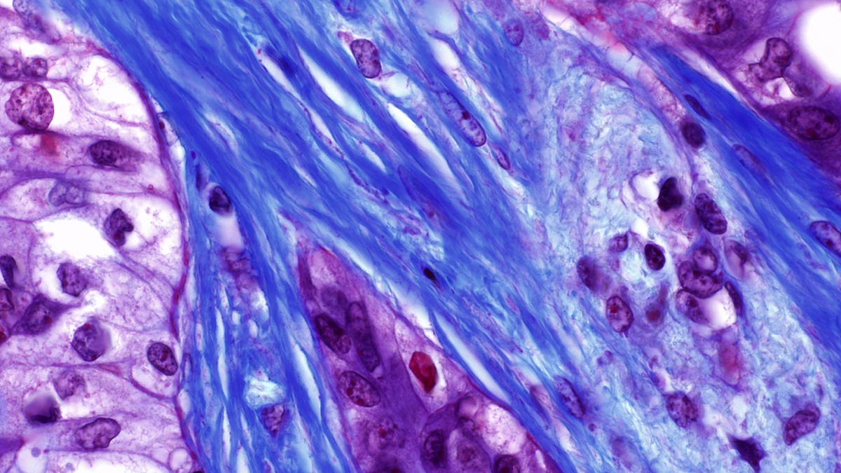
Conventional RNA-Seq generates an average summary of mRNA abundance across all of the cells in a sample. Recent research, however, has created a demand for higher resolution technologies capable of generating mRNA profiles at the level of single cells. In cancer biology, for example, there is an increasingly acute awareness that gene expression in the cells that make up malignant tumors is highly heterogeneous. This suggests that in order to understand how the cells work together to drive a tumor’s cancerous behavior, scientists need better methods for characterizing the entire ecology of cells of which it is made. Being able to quantify differences in gene expression cell by cell could be one valuable way to explore such complex environments and understand how they sustain malignancy.

Although several single cell RNA-seq technologies have been unveiled in the past two years, they are expensive to operate and are not optimized to produce data on the scale that is required for systems biology research, particularly in tissue specimens with limited numbers of cells. In a new paper just published in the journal Scientific Reports, however, researchers in the laboratory of Department of Systems Biology Assistant Professor Peter Sims describe a novel approach that offers several important advantages over other existing methods.

The new, automated platform builds on previous innovations in the Sims Lab to offer a cheap, efficient, and reliable way to simultaneously measure gene expression in thousands of individual cells from a single tissue sample. Using custom designed microwell plates, microfluidics, temperature control systems, and software, the technology captures, tags, and generates a readout of the complete transcriptome in each cell, providing robust data that can then be analyzed to distinguish functional diversity among the cells in the sample. Already, the technology is playing a key role in several research projects being conducted in the Department of Systems Biology and promises to become even more powerful as the field of single cell genomics continues to evolve.

An essay coauthored by Andrea Califano (Chair, Department of Systems Biology) and Gideon Bosker and published in the Wall Street Journal asks whether quantitative modeling could reveal the keys for turning cancer off. They write:

  • Disappointed with the slow pace of discovery and inclined to look for elegant, universal explanations for nature’s conundrums, many cancer researchers have increasingly been asking: Is there some sort of “Da Vinci Code” for cancer? And can we crack it using mathematics?

    Quantitative modeling has been extremely successful in disciplines as diverse as astronomy, physics, economics and computer science. Can “cancer quants”—scientists applying quantitative analyses to the landscape of cancer biology—find the answers we seek? And, if so, what would the new paradigm look like? 

The essay goes on to describe how computational methods developed in the Califano Lab are being tested in personalized N of 1 clinical trials to identify essential checkpoints in the molecular regulatory networks that sustain individual patients' tumors — as well as drugs capable of targeting them.

Click here to read the essay. (subscription may be required)

Yufeng Shen
Yufeng Shen's lab is interested in developing better computational methods for identifying rare genetic variants that increase disease risk.

On the surface, birth defects and cancer might not seem to have much in common. For some time, however, scientists have observed increased cancer risk among patients with certain developmental syndromes. One well-known example is seen in children with Noonan syndrome, who have an eightfold increased risk of developing leukemia. Recently, researchers studying the genetics of autism also observed mutations in PTEN, an important tumor suppressor gene. Although such findings have been largely isolated and anecdotal, they raise the tantalizing question of whether cancer and developmental disorders might be fundamentally linked.

According to a paper recently published in the journal Human Mutation, many of these similarities might not be just coincidental, but the result of shared genetic mutations. The study, led by Yufeng Shen, an Assistant Professor in the Columbia University Departments of Systems Biology and Biomedical Informatics, together with Wendy Chung, Kennedy Family Associate Professor of Pediatrics at Columbia University Medical Center, found that cancer-driving genes also make up more than a third of the risk genes for developmental disorders. Moreover, many of these genes appear to function through similar modes of action. The scientists suggest that this could make tumors “natural laboratories” for pinpointing and predicting the damaging effects of rare genetic alterations that cause developmental disorders.

“In comparison with cancer, there are relatively few patients with developmental disorders,” Shen explains, “For geneticists, this makes it hard to identify the risk genes solely based on statistical evidence of mutations from these patients. This study indicates that we should be able to use what we learn from cancer genetics — where much more data are available — to help in the interpretation of genetic data in developmental disorders.”

Master regulators of tumor homeostasis

In this rendering, master regulators of tumor homeostasis (white) integrate upstream genetic and epigenetic events (yellow) and regulate downstream genes (purple) responsible for implementing cancer programs such as proliferation and migration. CaST aims to develop systematic methods for identifying drugs capable of disrupting master regulator activity.

The Columbia University Department of Systems Biology has been named one of four inaugural centers in the National Cancer Institute’s (NCI) new Cancer Systems Biology Consortium. This five-year grant will support the creation of the Center for Cancer Systems Therapeutics (CaST), a collaborative research center that will investigate the general principles and functional mechanisms that enable malignant tumors to grow, evade treatment, induce disease progression, and develop drug resistance. Using this knowledge, the Center aims to identify new cancer treatments that target master regulators of tumor homeostasis.

CaST will build on previous accomplishments in the Department of Systems Biology and its Center for Multiscale Analysis of Genomic and Cellular Networks (MAGNet), which developed several key systems biology methods for characterizing the complex molecular machinery underlying cancer. At the same time, however, the new center constitutes a step forward, as it aims to move beyond a static understanding of cancer biology toward a time-dependent framework that can account for the dynamic, ever-changing nature of the disease. This more nuanced understanding could eventually enable scientists to better predict how individual tumors will change over time and in response to treatment.

Factors affecting protein activity
Following gene transcription and translation, a protein can undergo a variety of modifications that affect its activity. By analyzing downstream gene expression patterns in single tumors, VIPER can account for these changes to identify proteins that are critical to cancer cell survival.

In a paper just published in Nature Genetics, the laboratory of Andrea Califano introduces what it describes as the first method capable of analyzing a single tumor biopsy to systematically identify proteins that drive cancerous activity in individual patients. Based on knowledge gained by modeling networks of molecular interactions in the cell, their computational algorithm, called VIPER (Virtual Inference of Protein activity by Enriched Regulon analysis), offers a unique new strategy for understanding how cancer cells survive and for identifying personalized cancer therapeutics.

Developed by Mariano Alvarez as a research scientist in the Califano laboratory, VIPER has become one of the cornerstones of Columbia University’s precision medicine initiative. Its effectiveness in cancer diagnosis and treatment planning is currently being tested in a series of N-of-1 clinical trials, which analyze the unique molecular characteristics of individual patients’ tumors to identify drugs and drug combinations that will be most effective for them. If successful, it could soon become an important component of cancer care at Columbia University Medical Center.

According to Dr. Califano, “VIPER makes it possible to find actionable proteins in 100% of cancer patients, independent of their genetic mutations. It also enables us to track tumors as they progress or relapse to determine the most appropriate therapeutic approach at different points in the evolution of disease. So far, this method is looking extremely promising, and we are excited about its potential benefits in finding novel therapeutic strategies to treat cancer patients.”

Clonal evolution in GBM tumors
The researchers' model of tumor evolution indicates that different clonal lineages branch from a common ancestral cell and then diversify, independently causing aggressive tumor behavior at different stages of disease.

Glioblastoma multiforme (GBM) is the most common and most aggressive type of primary brain tumor in adults. Existing treatments against the disease are very limited in their effectiveness, meaning that in most patients tumors recur within a year. Once GBM returns, no beneficial therapeutics currently exist and prognosis is generally very poor.

To better understand how GBM evades treatment, an international team led by Antonio Iavarone and Raul Rabadan at the Columbia University Center for Topology of Cancer Evolution and Heterogeneity has been studying how the cellular composition of GBM tumors changes over the course of therapy. In a paper just published online by Nature Genetics, they provide the first sketch of the main routes of GBM tumor evolution during treatment, showing that different cellular clones within a tumor become dominant within specific tumor states. The study uncovers important general principles of tumor evolution, novel genetic markers of disease progression, and new potential therapeutic targets.


Andrea CalifanoAndrea Califano, the Clyde and Helen Wu Professor of Chemical Systems Biology and Chair of the Columbia University Department of Systems Biology, has been named a recipient of a National Cancer Institute Outstanding Investigator Award. The seven-year grant will support the development of systematic approaches for identifying the molecular factors that lead to cancer progression and to the emergence of drug resistance at the single-cell level. 

Breast cancer cells

A histological slide of cancerous breast tissue. The pink "riverways" are normal connective tissue while areas stained blue are cancer cells. (Source: National Cancer Institute)

Investigators at Columbia University Medical Center and the Icahn School of Medicine at Mount Sinai have discovered a molecular signaling mechanism that drives a specific type of highly aggressive breast cancer. As reported in a paper in Genes & Development, a team led by Jose Silva and Andrea Califano determined that the gene STAT3 is a master regulator of breast tumors lacking hormone receptors but testing positive for human epidermal growth receptor 2 (HR-/HER2+). The researchers also characterized a pathway including IL-6, JAK2, STAT3, and S100A8/9 — genes already known to play important roles within the immune response — as being essential for the survival of HR-/HER2+ cancer cells. Additional tests showed that disrupting this pathway severely limits the ability of these cells to survive.

These findings are particularly exciting because the pathway the researchers identified contains multiple targets for which known FDA-approved drugs exist. The paper reports that when these drugs were tested in disease models, the cancer cells showed a dramatic response, suggesting promising strategies for the treatment of the HR-/HER2+ cancer subtype. A clinical trial is now underway to investigate the effects of these approaches in humans.