2013 News

Imaging synapses

In a test of the Columbia Genome Center's high-content microscopy system, computational image analysis confirmed a high degree of colocalization of green fluorescent protein-labeled synapsin and the dye FM4-64. The researchers plan a high-throughput screen to identify fluroescent small molecules capable of targeting synapses.

Synapses mediate communication between neurons in the brain, making them critical components for neurological activity. Research has shown that synaptic loss and dysfunction play roles in a number of debilitating brain disorders — including Alzheimer’s disease, major depressive disorder, and autism — but currently no effective method exists for identifying and imaging individual synapses in living human brains. Being able to locate and quantify synapses in patients could greatly improve the diagnosis and monitoring of disease, and potentially offer new approaches for treatment.

Clarissa Waites, an assistant professor of pathology and cell biology at the Columbia University College of Physicians & Surgeons, and Dalibor Sames in the Columbia University Department of Chemistry, have recently embarked on a collaboration with the Columbia Genome Center High-Throughput Screening Facility with the goal of identifying small fluorescent molecules that can selectively localize to synapses. If successful, this project could for the first time provide a method for targeting and imaging synapses in the living human brain.

The magazine Genetic Engineering & Biotechnology News has published an article reporting on several talks given at the recent Department of Systems Biology Inaugural Symposium.

Author Richard A. Stein writes:

“Science is more than a body of knowledge, it’s a way of thinking,” remarked Carl Sagan, and probably his words were never more powerfully relevant than for portraying one of the newest biomedical fields, systems biology.

A recent symposium inaugurating the department of systems biology at Columbia University Medical Center comes at a very auspicious time, one in which biomedical sciences, chemistry, physics, engineering, bioinformatics, and computer sciences are converging to shape a vibrant new discipline.

The article includes summaries of presentations by Department of Systems Biology faculty members Barry Honig (protein-protein interactions), Saeed Tavazoie (gene expression dynamics), Virginia Cornish (synthetic chemistry using yeast), and Peter Sims (high-throughput single-cell measurement), as well as keynote speaker James Collins (antibiotics and the microbiome).

You can read the full article here.

Department of Systems Biology Symposium

On Thursday, October 17 more than 200 attendees filled the Hammer Health Sciences Center auditorium to celebrate the recent creation of the new Columbia University Department of Systems Biology. The event featured a keynote address by pioneering systems and synthetic biologist James Collins, as well as talks from more than a dozen Department faculty members and other collaborating investigators that spotlighted the wide range of research in computational and systems biology being pursued at Columbia. 

Models of Evolution In Charles Darwin's seminal treatise On the Origin of Species there is only one image, which visualizes evolution as following a branching pattern in which species diverge into lineages over time like the limbs on a tree. With the increasing availability of genomic data, scientists have attempted to understand evolution at the molecular level by using a similar phylogenetic paradigm, but as Department of Systems Biology Assistant Professor Raul Rabadan , MD/PhD student Joseph Chan, and Stanford University mathematician Gunnar Carlsson point out in a new paper published in the Proceedings of the National Academy of Sciences , it has a number of shortcomings when applied in this way. By developing a new mathematical approach based on a method called persistent homology, the researchers produced several insights into viral evolution that could not be found using other means.

Researchers in the Columbia University Department of Systems Biology and Herbert Irving Comprehensive Cancer Center have determined that measuring the expression levels of three genes associated with aging can be used to predict the aggressiveness of seemingly low-risk prostate cancer. Use of this three-gene biomarker, in conjunction with existing cancer-staging tests, could help physicians better determine which men with early prostate cancer can be safely followed with “active surveillance” and spared the risks of prostate removal or other invasive treatment. The findings were published today in the online edition of Science Translational Medicine.

More than 200,000 new cases of prostate cancer are diagnosed each year in the U.S. “Most of these cancers are slow growing and will remain so, and thus they do not require treatment,” said study leader Cory Abate-Shen, Michael and Stella Chernow Professor of Urological Oncology at Columbia University Medical Center (CUMC). “The problem is that, with existing tests, we cannot identify the small percentage of slow-growing tumors that will eventually become aggressive and spread beyond the prostate. The three-gene biomarker could take much of the guesswork out of the diagnostic process and ensure that patients are neither overtreated nor undertreated.”

Rabadan, Nature Genetics

An analysis of all gene mutations in nearly 140 brain tumors has uncovered most of the genes responsible for driving glioblastoma. The analysis found 18 new driver genes (labeled red), never before implicated in glioblastoma and correctly identified the 15 previously known driver genes (labeled blue). The graphs show mutated genes that are commonly found in varying numbers in glioblastoma (left), that frequently contain insertions (middle), and that frequently contain deletions (right). Genes represented by blue dots in the graphs were statistically most likely to be driver genes.

A team of Columbia University Medical Center researchers has identified 18 new genes responsible for driving glioblastoma multiforme, the most common—and most aggressive—form of brain cancer in adults. The study was published August 5, 2013, in the journal Nature Genetics.

The Columbia team used a combination of high-throughput DNA sequencing and a new method of statistical analysis developed by co-author Raul Rabadan, an assistant professor in the Department of Systems Biology, to generate a short list of candidate gene mutations that were highly likely to drive cancer, as opposed to mutations that have no effect.

Considering these results along with a previous study this group conducted, Rabadan and collaborators Antonio Iavarone and Anna Lasorella point out that approximately 15% of glioblastomas could now be targeted with drugs that have already been approved by the FDA. As Lasorella remarks in an article for the CUMC Newsroom, “There is no reason why these patients couldn’t receive these drugs now in clinical trials.”

Reposted from the Columbia University Medical Center Newsroom. Find the original article here .
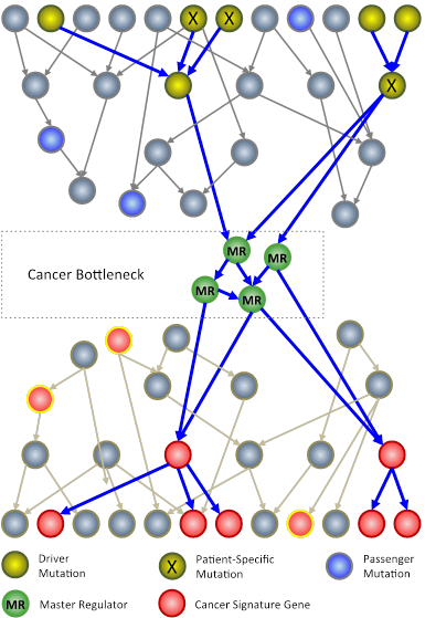
Cancer bottlenecks
In an N-of-1 study, researchers at Columbia University use techniques from systems biology to analyze genomic information from an individual patient’s tumor. The goal is to identify key genes, called master regulators  (green circles), which, while not mutated, are nonetheless necessary for the survival of cancer cells. 

Columbia University Medical Center (CUMC) researchers are developing a new approach to cancer clinical trials, in which therapies are designed and tested one patient at a time. The patient’s tumor is “reverse engineered” to determine its unique genetic characteristics and to identify existing U.S. Food and Drug Administration (FDA)-approved drugs that may target them.

Rather than focusing on the usual mutated genes, only a very small number of which can be used to guide successful therapeutic strategies, the method analyzes the regulatory logic of the cell to identify genes and gene pairs that are critical for the survival of the tumor but are not critical for normal cells. FDA-approved drugs that inhibit these genes are then tested in a mouse model of the patient’s tumor and, if successful, considered as potential therapeutic agents for the patient — a journey from bedside to bench and back again that takes about six to nine months.

“We are taking a rather different approach to tailor therapy to the individual cancer patient,” said principal investigator Andrea Califano, PhD, Clyde and Helen Wu Professor of Chemical Systems Biology and chair of CUMC’s new Department of Systems Biology. “If we have learned one thing about this disease, it’s that it has tremendous heterogeneity both across patients and within individual patients. When we expect different patients with the same tumor subtype or different cells within the same tumor to respond the same way to a treatment, we make a huge simplification. Yet this is how clinical studies are currently conducted. To address this problem, we are trying to understand how tumors are regulated one at a time. Eventually, we hope to be able to treat patients not on an individual basis, but based on common vulnerabilities of the cancer cellular machinery, of which genetic mutations are only indirect evidence. Genetic alterations are clearly responsible for tumorigenesis but control points in molecular networks may be better therapeutic targets.”

Columbia University Department of Systems Biology

Attendees at the June 2013 Columbia University Department of Systems Biology retreat.

Effective July 1, 2013, the Columbia Initiative in Systems Biology is now the Columbia University Department of Systems Biology. Approved by a vote of the University Trustees, this step recognizes the growth in systems biology research and education that has taken place at Columbia, and formally establishes this emerging discipline as a major area for research at the university.

As Andrea Califano, chair of the new department, explained, "This achievement testifies to the dedicated community that has been gathering at Columbia over the past decade around the field of systems biology. We have witnessed the emergence of a compelling scientific agenda that combines innovative experimental and quantitative methods to address important biological and biomedical questions in ways that were unimaginable just a few years ago. It is very exciting to see Columbia take this step because systems biology is paving the way toward new, more rational approaches in basic and translational research."

viSNE

viSNE reveals the progression of cancer in a sample of cells taken from a patient with acute myeloid leukemia. Cells are colored according to intensity of expression of the indicated cell markers, enabling the comparison of expression patterns before and after relapse. For example, Fit3 is expressed primarily in the diagnosis sample, while CD34 emerges in the relapse sample.

Researchers in the Columbia Initiative in Systems Biology have developed a computational method that enables scientists to visualize and interpret high-dimensional data produced by single-cell measurement technologies such as mass cytometry. The method, called viSNE (visual interactive Stochastic Neighbor Embedding), has just been published in the online edition of Nature Biotechnology. It has particular relevance to cancer research and therapeutics. As Columbia University Medical Center reports:

Researchers now understand that cancer within an individual can harbor subpopulations of cells with different molecular characteristics. Groups of cells may behave differently from one another, including in how they respond to treatment. The ability to study single cells, as well as to identify and characterize subpopulations of cancerous cells within an individual, could lead to more precise methods of diagnosis and treatment.

“Our method not only will allow scientists to explore the heterogeneity of cancer cells and to characterize drug-resistant cancer cells, but also will allow physicians to track tumor progression, identify drug-resistant cancer cells, and detect minute quantities of cancer cells that increase the risk of relapse,” said co-senior author Dana Pe’er, associate professor of biological sciences and systems biology at Columbia.

Figure

Tumor-induced mRNA expression changes for individual biochemical reactions in central metabolism. 

A large study analyzing gene expression data from 22 cancer types has identified a broad spectrum of metabolic expression changes associated with cancer. The analysis, led by Dennis Vitkup, first author Jie Hu, a postdoctoral research scientist in the Vitkup lab, with a multi-institutional group of collaborators, also identified hundreds of potential drug targets that could cut off a tumor’s fuel supply or interfere with its ability to synthesize essential elements necessary for tumor growth. The study has just been published in the online edition of Nature Biotechnology .

As Columbia University Medical Center reports:

The results should ramp up research into drugs that interfere with cancer metabolism, a field that dominated cancer research in the early 20th century and has recently undergone a renaissance.

Attractor Metagenes - DREAM7

Team Attractor Metagenes receives its award at the DREAM7 Conference. Gustavo Stolovitzky (IBM Research), Adam Margolis (Sage Bionetworks), Dimitris Anastassiou, Tai-Hsien Ou Yang, Wei-Yi Cheng, Stephen Friend (Sage Bionetworks), Erhan Bilal (IBM Research)

The team of Professor Dimitris Anastassiou and graduate students Wei-Yi Cheng and Tai-Hsien Ou Yang has been recognized as the best performer in the Sage Bionetworks – DREAM Breast Cancer Prognosis Challenge. This challenge, one of four organized as part of the seventh Dialogue for Reverse Engineering Assessments and Methods (DREAM7), was designed to assess the ability of participants’ computational models to predict breast cancer survival using patient clinical information and molecular profiling data. As a reward for this accomplishment, the journal Science Translational Medicine has just published a paper from the Anastassiou lab describing their model. It is also the journal’s cover theme for this issue, which includes a second article describing the Challenge.

The Columbia University researchers based their DREAM entry on previous work to identify what they call “attractor metagenes,” sets of strongly co-expressed genes that they have found to be present with very little variation in many cancer types. Moreover, these metagenes appear to be associated with specific attributes of cancer including chromosomal instability, epithelial-mesenchymal transition, and a lymphocyte-specific immune response. As Wei-Yi Cheng comments in Sage Synapse, “We like to think of these three main attractor metagenes as representing three key ‘bioinformatic hallmarks of cancer,’ reflecting the ability of cancer cells to divide uncontrollably and invade surrounding tissues, and the ability of the organism to recruit a particular type of immune response to fight the disease.”

Harris WangHarris Wang has joined Columbia University Medical Center as an Assistant Professor in the Columbia Initiative in Systems Biology and the Department of Pathology and Cell Biology. His research focuses on understanding the evolution of the ecosystems that develop within heterogeneous microbial communities. Using approaches from genome engineering, DNA synthesis, and next-generation sequencing, he studies how genomes in microbial populations form, maintain themselves, and change over time, both within and across microbial communities. His goal is to use synthetic biology approaches to engineer ecologies of microbial populations, such as those found in the gut and elsewhere in the human body, in ways that could improve human health.

Searches for hyperglycemia-related terms

Percentage of users in each of the three user groups searching for hyperglycemia-related terms, computed per week over 12 months of search log data. Background refers to the fraction of all searchers who search for hyperglycemia-related symptoms or terminology independent of the presence of the drugs in the users’ search histories.

Although the US Food and Drug Organization and other agencies collect and analyze reports on adverse drug effects, alerts for single drugs and drug-drug interactions are often delayed due to the time it takes to accumulate evidence. Columbia University Department of Systems Biology faculty member Nicholas Tatonetti, in collaboration with investigators at Stanford University and Microsoft Research, hypothesized that Internet users can provide early clues of adverse drug events as they seek information on the web concerning symptoms they are experiencing. A new paper explains their results.

As a test, Tatonetti and colleagues asked whether it would be possible to detect evidence of an interaction between the antidepressant paroxetine and the anti-cholesterol drug pravastatin by analyzing web search logs from 2010. As a postfoc at Stanford, Tatonetti and colleagues used a data mining algorithm to analyze FDA adverse event reporting records, and retroactively found this combination to be associated with hyperglycemia (high blood sugar) in some patients. In this new project, the researchers analyzed the search logs of millions of Internet users from a period before the above association was identified to see how often they entered search terms related to hyperglycemia and to one or both medications under investigation. (Participants in this study opted in by voluntarily installing a web browser extension that tracked their activity anonymously.)

High-throughput screening’s ability to perform thousands of experiments efficiently and under carefully controlled conditions has made it an important tool for basic and translational biological research. At Columbia University, the JP Sulzberger Columbia Genome Center and the Chemical Probe Synthesis Facility provide a flexible platform for researchers interested in applying high-throughput experimentation in their work. On December 17, 2012, the Genome Center hosted a symposium to spotlight its capabilities in high-throughput screening, to explain the important role that synthetic chemistry plays in high-throughput screening, and to describe some recent research projects at Columbia that have utilized these tools.

As High-Throughput Screening Core Scientific Director Charles Karan explained, the Genome Center operates a suite of advanced technologies for automated liquid handling, robotic assay implementation, and high-throughput, high-content microscopy. The Genome Center also offers Columbia University researchers access to several large collections for conducting high-throughput screens. These include the Columbia Cell Line Encyclopedia, which includes 850 cancer cell lines collected from around the world, as well as a chemical diversity library curated by researchers in the Chemical Probe Synthesis Facility. This “tool chest” gives Columbia investigators access to a pre-selected set of compounds that have been predicted to result in the highest quality potential hits. Karan also reported that the Genome Center recently negotiated an arrangement with Sigma Aldrich to give access to the company’s shRNA clones to researchers at Columbia at greatly discounted rates.

Itsik Pe'erItsik Pe'er, an Associate Professor in the Department of Computer Science and member of the Columbia Initiative in Systems Biology, is using mathematics and computer analytics to identify the genetic makeup of the founding Ashkenazi Jews. By analyzing the full DNA sequences of hundreds of their descendants in the New York City area and comparing them to reference sets of non-Ashkenazi DNA, his goal is to identify Ashkenazi-specific genetic mutations associated with diseases such as Tay-Sachs, Crohn's, and Parkinson's disease. As a new article in Columbia News explains:

By examining similarities in DNA segments shared by large numbers of related individuals, his lab developed statistical models that allow him to make generalizations about entire populations. The mix of genes that every child inherits from each parent travels in long sequences of code that remain together and are remarkably consistent from one generation to the next.

"The size of the gene chunks gets smaller with each generation, but they diminish at a consistent and predictable rate. As a result, Pe’er can use his models to determine distant relationships shared by two individuals by measuring the length of their common DNA segments."

Read the complete article here.

The cell-of-origin model in cancer biology suggests that some tumors are more aggressive than others because of differences in the cell lineages from which they arise. In the prostate gland, there are three types of epithelial stem cell — luminal cells, basal cells, and rare neuroendocrine cells. There has been some discrepancy in the scientific literature, however, about whether luminal cells, basal cells, or both can act as a cell of tumor origin.

In a paper published online in the journal Nature Cell Biology, researchers in the laboratories of Columbia University Department of Systems Biology members Michael Shen, Andrea Califano, and Cory Abate-Shen undertook a comprehensive analysis of prostate basal cell properties in mouse models. They used a technique called genetic linkage marking to study an identical cell population in multiple assays of stem cell function.

The studies showed that discrepancies in the published literature arise because basal stem cell properties can change when studied outside their endogenous tissue microenvironment; that is, in ex vivo cell culture and tissue grafting assays. To avoid this problem, they suggest, genetic lineage tracing in vivo should be considered the gold standard for identifying physiologically relevant stem cells.